

Clinical Subject Page
Sarcoidosis
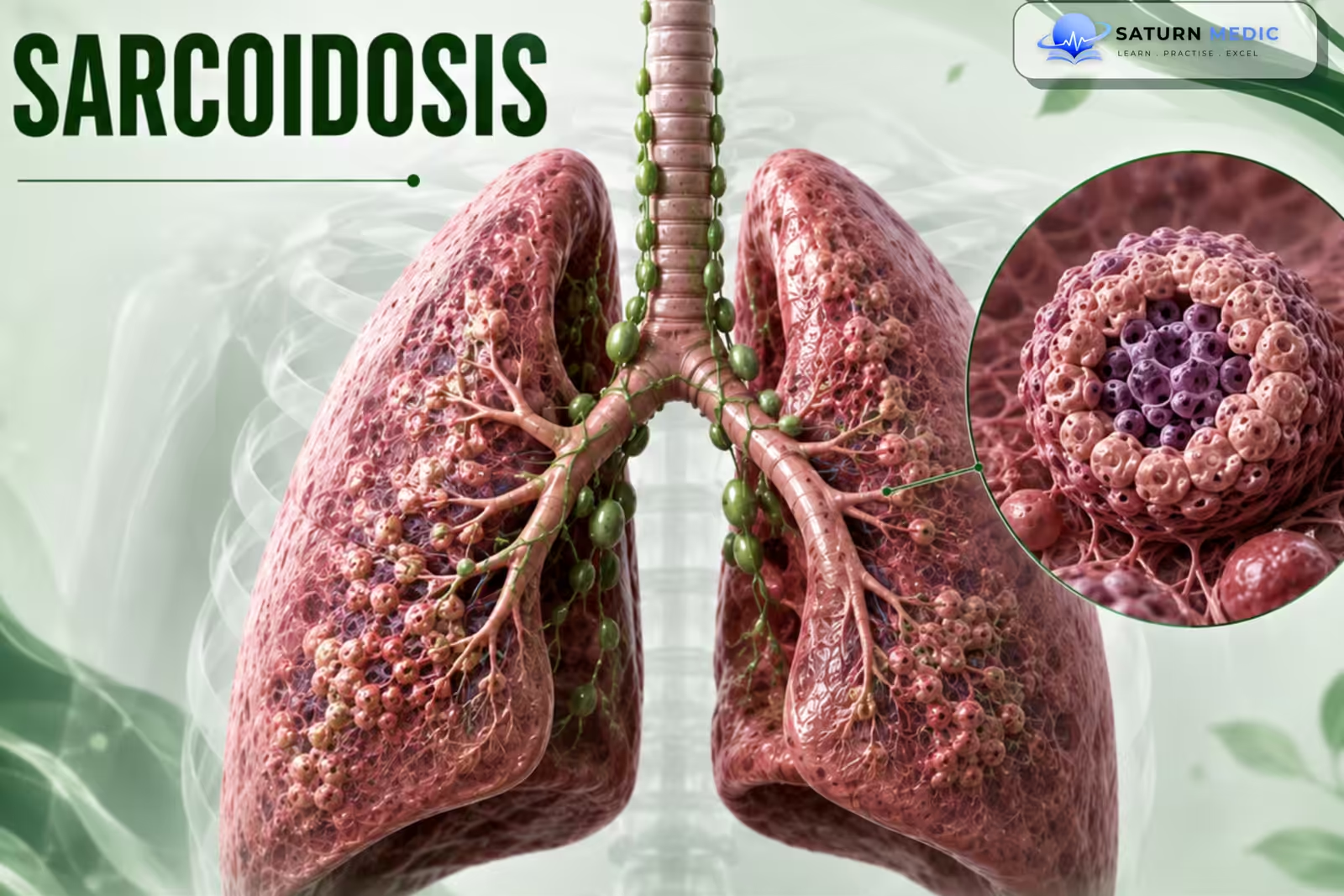
Sarcoidosis is a multisystem inflammatory disease characterized by the formation of noncaseating granulomas, most commonly affecting the lungs and intrathoracic lymph nodes.
Also called
ICD-10
Specialty
Onset
Reviewed
-
OverviewOverview
-
Etiology & Risk FactorsEtiology & Risk Factors
-
PathophysiologyPathophysiology
-
Clinical PresentationClinical Presentation
-
History TakingHistory Taking
-
Physical ExaminationPhysical Examination
-
InvestigationsInvestigations
-
DiagnosisDiagnosis
-
ManagementManagement
-
ComplicationsComplications
-
PrognosisPrognosis
-
Key Points / Clinical PearlsKey Points / Clinical Pearls
Overview
Sarcoidosis can affect almost any organ, but most commonly involves the:
- Lungs
- Intrathoracic lymph nodes
- Skin
- Eyes
The disease results from an abnormal immune response to an unknown trigger in genetically susceptible individuals.
Etiology & Risk Factors
Etiology
The exact cause is unknown.
Sarcoidosis probably develops through an interaction between:
-
Genetic susceptibility
-
Environmental or infectious triggers
-
Abnormal immune response
Risk Factors
-
Young and middle adulthood
-
Family history of sarcoidosis
-
Certain genetic backgrounds
-
Exposure to specific environmental or occupational agents
Important Note
Sarcoidosis is not an infectious disease, although infectious agents may act as possible triggers.
Pathophysiology
Unknown environmental or infectious trigger
⬇
Abnormal immune activation
⬇
Activation of T lymphocytes and macrophages
⬇
Accumulation of inflammatory cells
⬇
Formation of noncaseating granulomas
⬇
Organ inflammation and dysfunction
⬇
Granuloma resolution OR chronic inflammation
⬇
Fibrosis and permanent organ damage in some patients
Key Concept
The hallmark of sarcoidosis is the noncaseating granuloma.
Clinical Presentation
General Symptoms
-
Fatigue
-
Fever
-
Weight loss
-
Night sweats
Pulmonary Features
-
Dry cough
-
Progressive dyspnea
-
Chest discomfort
-
Wheezing
Skin Features
-
Erythema nodosum
-
Lupus pernio
-
Papules or plaques
Eye Features
-
Uveitis
-
Eye pain
-
Redness
-
Photophobia
-
Visual disturbance
Other Features
-
-
Peripheral lymphadenopathy
-
Arthralgia or arthritis
-
Cardiac arrhythmias
-
Neurological symptoms
-
Hypercalcemia
-
History Taking
-Ask about:
Dry cough?
Shortness of breath?
Chest discomfort?
Fever or night sweats?
Weight loss?
Fatigue?
Skin rash or painful leg nodules?
Eye pain or redness?
Visual changes?
Joint pain or swelling?
Family history of sarcoidosis?
Physical Examination
-General Examination
Look for:
- Peripheral lymphadenopathy
- Erythema nodosum
- Lupus pernio
- Other skin lesions
Chest Examination
May show:
- Fine crackles
- Wheezing
The chest examination may also be normal.
-Eye Examination
Look for:
- Eye redness
- Signs of uveitis
-Other Examination
Look for:
-
- Joint swelling
- Hepatomegaly
- Splenomegaly
- Neurological abnormalities
- Signs of cardiac involvement
Investigations
-Chest Imaging — Key Investigation
Chest X-ray
May show:
-
Bilateral hilar lymphadenopathy
-
Pulmonary infiltrates
-
Pulmonary fibrosis
Chest CT
Provides better assessment of:
-
Lymphadenopathy
-
Lung nodules
-
Parenchymal disease
-
Fibrosis
-Pulmonary Function Tests
May show:
-
Restrictive pattern
-
Obstructive pattern
-
Reduced DLCO
-
Normal results
-Blood Tests
May include:
-
CBC
-
Renal function
-
Liver function
-
Serum calcium
-
Vitamin D assessment when indicated
-ECG
Used to screen for possible cardiac involvement.
Tissue Biopsy
May show:
-
Noncaseating granulomas
Important Note
Serum ACE levels are neither sensitive nor specific enough to confirm or exclude sarcoidosis.
Diagnosis
Sarcoidosis · Diagnostic Criteria & Stepwise Approach
Biopsy of the most accessible lesion is preferred. Löfgren syndrome (erythema nodosum + bilateral hilar lymphadenopathy + arthralgia) may be diagnosed clinically without biopsy.
| CXR Stage | Radiological Findings | Spontaneous Remission Rate |
|---|---|---|
| Stage 0 | Normal chest radiograph — extrapulmonary disease only | N/A |
| Stage I | Bilateral hilar lymphadenopathy (BHL) alone — no parenchymal involvement | 55–90% spontaneous remission |
| Stage II | BHL + pulmonary parenchymal infiltrates | 40–70% spontaneous remission |
| Stage III | Pulmonary infiltrates without BHL | 10–20% spontaneous remission |
| Stage IV | Pulmonary fibrosis, honeycombing, bullae, traction bronchiectasis — end-stage | Irreversible — no remission |
Related Topics
Management
Sarcoidosis · Treatment
Systemic corticosteroids remain the backbone of therapy; steroid-sparing agents reduce long-term steroid toxicity. There is no cure.
| Drug / Treatment | Line | Dose / Notes |
|---|---|---|
| Prednisolone | First-line | 0.5 mg/kg/day (typically 20–40 mg/day). Taper after 4–6 weeks; minimum 12-month course. Use bone protection (calcium, vitamin D, bisphosphonates). Monitor glucose, BP, eyes (cataracts/glaucoma). |
| Methotrexate | Steroid-sparing | 7.5–15 mg/week orally + folic acid 5 mg once weekly (day after MTX). Most widely used second-line agent. Allows steroid dose reduction. Monitor FBC, LFTs monthly. Avoid in pregnancy. |
| Azathioprine | Steroid-sparing | 1–2 mg/kg/day. Check TPMT enzyme level before starting. Alternative to methotrexate. Monitor FBC and LFTs. Used in hepatic or neurological sarcoidosis. |
| Hydroxychloroquine | Steroid-sparing | 200–400 mg/day. Preferred for skin, hypercalcaemia, and constitutional symptoms (fatigue). Less effective for pulmonary disease. Annual retinal monitoring required (≥5 years of use). |
| Infliximab (anti-TNFα) | Third-line / Refractory | 3–5 mg/kg IV infusion at 0, 2, 6 weeks then every 4–8 weeks. Effective in refractory/chronic sarcoidosis. Screen for TB/hepatitis B before use. Best evidence for pulmonary and cutaneous disease. |
| Adalimumab | Third-line / Refractory | Subcutaneous anti-TNF; alternative to infliximab for refractory disease. Particularly used in ocular and cutaneous sarcoidosis. |
| Topical/Local Corticosteroids | Organ-specific | Topical eye drops (prednisolone/dexamethasone) for anterior uveitis — first-line before systemic. Inhaled corticosteroids for cough/bronchial sarcoidosis — limited systemic absorption. |
| NSAIDs | Symptomatic | For Löfgren syndrome arthralgia and erythema nodosum. Avoid systemic corticosteroids unless refractory in Löfgren. Short-course only. |
| ICD / Pacemaker | Cardiac sarcoidosis | Complete heart block → permanent pacemaker. Sustained VT/LVEF <35% → ICD. Systemic steroids for active cardiac inflammation (40 mg/day prednisolone). High mortality without treatment. |
| Lung Transplantation | End-stage (Stage IV) | For Stage IV pulmonary fibrosis with severe impairment. Sarcoidosis can recur in transplanted lungs (rare). Refer when FVC <50% or O2-dependent. |
Complications
- Pulmonary fibrosis
- Chronic respiratory failure
- Pulmonary hypertension
- Vision loss
- Cardiac arrhythmias
- Heart block
- Heart failure
- Sudden cardiac death
- Neurological damage
- Hypercalcemia
- Kidney stones
- Chronic organ dysfunction
Prognosis
- The course is highly variable.
- Many patients experience spontaneous remission.
- Others develop chronic or progressive disease.
- Prognosis is worse with:
- Pulmonary fibrosis
- Pulmonary hypertension
- Cardiac involvement
- Neurological involvement
- Chronic progressive disease
- Early recognition of serious organ involvement improves outcomes.
Key Points / Clinical Pearls
- Sarcoidosis is a multisystem granulomatous disease.
- The lungs and intrathoracic lymph nodes are most commonly affected.
- Noncaseating granulomas are the pathological hallmark.
- Bilateral hilar lymphadenopathy is a classic imaging finding.
- Always exclude infections and other causes of granulomas.
- Serum ACE does not confirm or exclude the diagnosis.
- Not every patient requires treatment.
- Corticosteroids are the main initial treatment for significant disease.
- Always consider eye, cardiac, and neurological involvement.
- National Heart, Lung, and Blood Institute (NIH). Sarcoidosis.
- MedlinePlus, National Library of Medicine (NIH). Sarcoidosis: Medical Encyclopedia.
- Crouser ED, Maier LA, Wilson KC, et al. Diagnosis and Detection of Sarcoidosis: An Official American Thoracic Society Clinical Practice Guideline. Am J Respir Crit Care Med. 2020. PMID: 32293205.