

Clinical Subject Page
Iron Deficiency Anemia (IDA)
Iron Deficiency Anemia (IDA) is the most common nutritional deficiency and the most common cause of anemia worldwide. It develops when iron stores become insufficient to support normal hemoglobin synthesis, resulting in reduced oxygen-carrying capacity of the blood.
Also called
ICD-10
Specialty
Onset
Reviewed
-
OverviewOverview
-
Etiology & Risk FactorsEtiology & Risk Factors
-
PathophysiologyPathophysiology
-
Clinical PresentationClinical Presentation
-
History TakingHistory Taking
-
Physical ExaminationPhysical Examination
-
InvestigationsInvestigations
-
DiagnosisDiagnosis
-
ManagementManagement
-
ComplicationsComplications
-
PrognosisPrognosis
-
Key Points / Clinical PearlsKey Points / Clinical Pearls
Overview
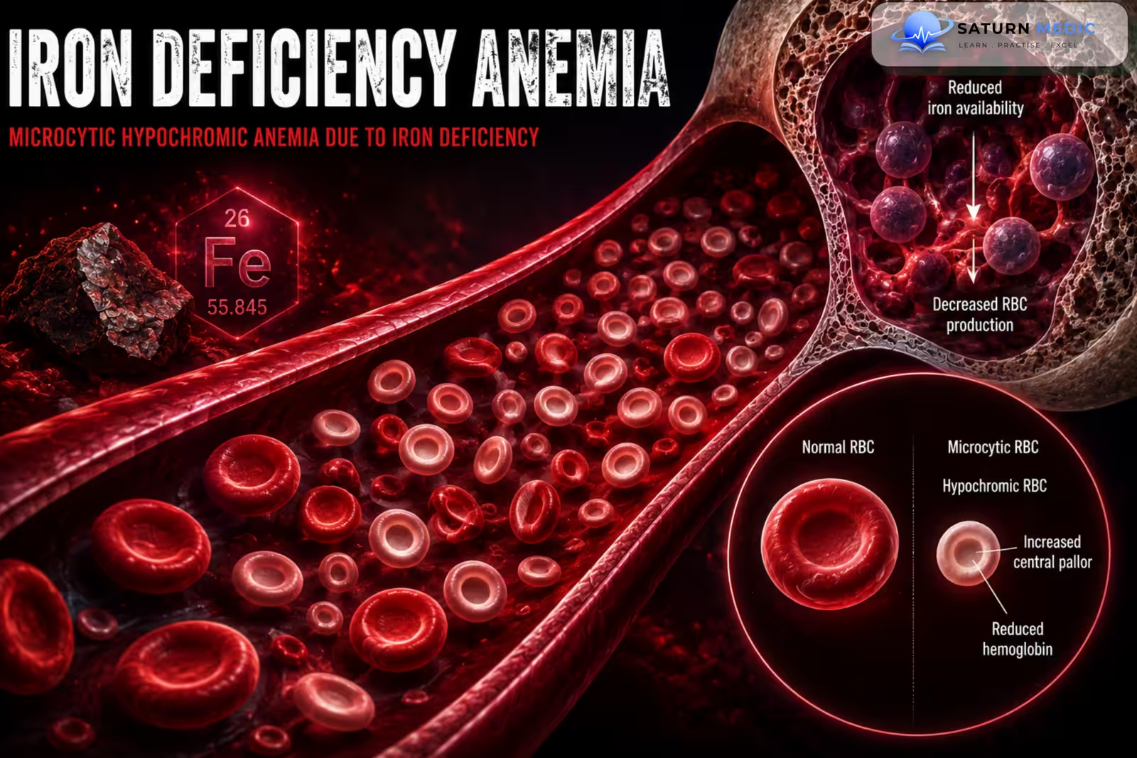
Iron Deficiency Anemia (IDA) occurs when iron availability is insufficient for hemoglobin production. The main causes are chronic blood loss, inadequate dietary intake, increased physiological requirements, and impaired iron absorption. It typically produces a microcytic, hypochromic anemia with low ferritin and low serum iron.
In adults, particularly men and postmenopausal women, Iron Deficiency Anemia (IDA) should be considered a potential sign of occult gastrointestinal blood loss until proven otherwise.
Etiology & Risk Factors
Etiology & Risk Factors
Aetiology
1. Chronic Blood Loss
Gastrointestinal bleeding
Peptic ulcer disease
Gastric or colorectal cancer
Inflammatory bowel disease
Hemorrhoids
Gastrointestinal polyps
Menorrhagia
Recurrent epistaxis
Frequent blood donation
2. Increased Iron Requirements
Pregnancy
Infancy
Childhood
Adolescence
3. Reduced Iron Intake
Poor dietary intake
Malnutrition
Restrictive diets
4. Impaired Iron Absorption
Celiac disease
Atrophic gastritis
H. pylori infection
Previous gastric or bariatric surgery
Inflammatory bowel disease
Long-term acid suppression
Risk Factors
Menstruation
Pregnancy
Older age
Vegetarian or vegan diet
Chronic gastrointestinal disease
Previous gastrointestinal surgery
Chronic NSAID use
Gastrointestinal malignancy
Frequent blood donation
Pathophysiology
Reduced iron intake, increased iron requirements, chronic blood loss, or impaired absorption → depletion of iron stores → decreased serum ferritin → reduced iron availability for erythropoiesis → decreased hemoglobin synthesis → production of smaller and paler red blood cells → microcytic hypochromic anemia → reduced oxygen delivery to tissues → compensatory increase in cardiac output → symptoms of anemia and tissue hypoxia.
Clinical Presentation
-Symptoms
Fatigue
Weakness
Reduced exercise tolerance
Shortness of breath on exertion
Dizziness
Headache
Palpitations
Poor concentration
Irritability
-Signs
Pallor
Tachycardia
Brittle nails
Koilonychia (spoon-shaped nails)
Glossitis
Angular cheilitis
Hair loss
-Specific Features of Iron Deficiency
Pica
Pagophagia (ice craving)
Restless legs syndrome
History Taking
-Ask about:
- Fatigue and weakness
- Shortness of breath
- Dizziness
- Palpitations
- Dietary intake
- Menstrual history
- Pregnancy
- Gastrointestinal symptoms
- Melena
- Hematochezia
- Abdominal pain
- Change in bowel habits
- Weight loss
- Dysphagia
- NSAID use
Physical Examination
General Examination
Look for:
Pallor
Tachycardia
Signs of heart failure in severe cases
Weight loss
General nutritional status
Specific Examination
Assess for:
Koilonychia
Glossitis
Angular cheilitis
Brittle hair
Hair loss
Lymphadenopathy
Abdominal Examination
Look for:
Abdominal masses
Hepatosplenomegaly
Abdominal tenderness
Rectal Examination
Consider when gastrointestinal bleeding or colorectal pathology is suspected.
Investigations
Complete Blood Count (CBC)
Typical findings:
Low hemoglobin
Low hematocrit
Low MCV
Low MCH
Low MCHC
Increased RDW
Iron Studies
Serum Ferritin
Low ferritin is the most specific laboratory finding for iron deficiency.
Ferritin may be falsely normal or elevated in inflammation because it is an acute-phase reactant.
Serum Iron
Low
Total Iron-Binding Capacity (TIBC)
Increased
Transferrin Saturation
Reduced
Peripheral Blood Film
May show:
Microcytosis
Hypochromia
Anisocytosis
Poikilocytosis
Pencil cells
Reticulocyte Count
Usually low or inappropriately normal before treatment
Increases after effective iron replacement
Additional Investigations to Identify the Cause
Depending on age, sex, and clinical presentation:
Stool testing for occult blood
Upper gastrointestinal endoscopy
Colonoscopy
Celiac disease screening
H. pylori testing
Gynecological evaluation in women with abnormal uterine bleeding
Urinalysis for urinary blood loss
Diagnosis
Diagnosis is based on:
- Low hemoglobin
- Microcytic or sometimes initially normocytic anemia
- Low ferritin
- Low serum iron
- Increased TIBC
- Low transferrin saturation
- Clinical evidence of iron deficiency
The underlying cause must always be investigated.
Management
1. Treat the Underlying Cause
Control gastrointestinal bleeding
Treat peptic ulcer disease
Treat H. pylori infection
Treat celiac disease
Manage heavy menstrual bleeding
Treat gastrointestinal malignancy if present
Stop or modify offending medications when appropriate
2. Oral Iron Therapy
First-line treatment for most patients:
Oral ferrous sulfate
Ferrous fumarate
Ferrous gluconate
Treatment may be given once daily or on alternate days depending on tolerance and clinical circumstances.
Common adverse effects:
Nausea
Constipation
Abdominal discomfort
Dark stools
3. Intravenous Iron
Consider when:
Oral iron is not tolerated
Oral iron is ineffective
Malabsorption is present
Significant ongoing blood loss exists
Rapid iron replacement is needed
Chronic kidney disease or inflammatory conditions limit response
4. Blood Transfusion
Reserved for:
Severe symptomatic anemia
Hemodynamic instability
Active major bleeding
Severe tissue hypoxia
Monitoring
Monitor hemoglobin response
Check reticulocyte response when appropriate
Repeat iron studies after treatment
Continue iron therapy after hemoglobin normalization to replenish iron stores
Complications
- Severe fatigue
- Reduced exercise capacity
- Cognitive impairment
- Poor school or work performance
- Restless legs syndrome
- Cardiovascular strain
- Heart failure in severe prolonged cases
- Pregnancy complications
- Increased susceptibility to complications from the underlying cause
Prognosis
The prognosis of Iron Deficiency Anemia (IDA) is generally excellent when the underlying cause is identified and treated. Hemoglobin usually improves with adequate iron replacement, but replenishment of total body iron stores takes longer. Persistent or recurrent Iron Deficiency Anemia (IDA) requires reassessment for ongoing blood loss, malabsorption, poor adherence, or an incorrect diagnosis.
Key Points / Clinical Pearls
- Iron Deficiency Anemia (IDA) is the most common cause of anemia worldwide.
- The main causes are chronic blood loss, increased requirements, poor intake, and malabsorption.
- It is typically a microcytic, hypochromic anemia.
- Low ferritin is the key laboratory marker of depleted iron stores.
- Serum iron is low and TIBC is usually increased.
In adult men and postmenopausal women, investigate for occult gastrointestinal blood loss. - Oral iron is first-line for most patients.
IV iron is useful when oral iron is ineffective, poorly tolerated, or contraindicated. - Blood transfusion is reserved for severe symptomatic anemia or significant instability.
- Always identify and treat the underlying cause of Iron Deficiency Anemia (IDA).
- National Center for Biotechnology Information (NIH). Iron Deficiency and Microcytic Hypochromic Anemia, StatPearls.
- American Society of Hematology (ASH). ASH Clinical Practice Guidelines on Iron Deficiency.
- National Center for Biotechnology Information (NIH). Anemia, StatPearls.
- World Health Organization. Anaemia: Fact Sheet.
- MedlinePlus, National Library of Medicine (NIH). Iron-Deficiency Anemia: Health Topic.