

Clinical Subject Page
Sickle Cell Anemia (SCA)
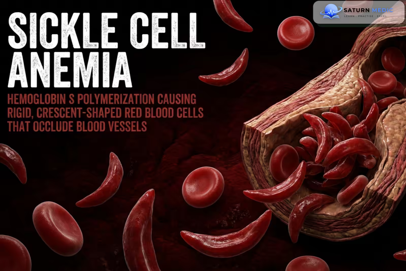
Sickle Cell Anemia (SCA) is an inherited hemoglobin disorder caused by a mutation in the β-globin gene, resulting in production of abnormal hemoglobin S (HbS). Under low-oxygen conditions, HbS polymerizes, causing red blood cells to become rigid and sickle-shaped, leading to chronic hemolytic anemia and recurrent vaso-occlusive episodes.
Also called
ICD-10
Specialty
Onset
Reviewed
-
OverviewOverview
-
Etiology & Risk FactorsEtiology & Risk Factors
-
PathophysiologyPathophysiology
-
Clinical PresentationClinical Presentation
-
History TakingHistory Taking
-
Physical ExaminationPhysical Examination
-
InvestigationsInvestigations
-
DiagnosisDiagnosis
-
ManagementManagement
-
ComplicationsComplications
-
PrognosisPrognosis
-
Key Points / Clinical PearlsKey Points / Clinical Pearls
Overview
Sickle Cell Anemia (SCA) is an autosomal recessive inherited disorder caused by homozygous HbS (HbSS). Sickled red blood cells have reduced deformability and increased adherence to vascular endothelium, causing microvascular obstruction and tissue ischemia.
The disease produces two major problems: chronic hemolysis, causing anemia and jaundice, and vaso-occlusion, causing recurrent painful crises and progressive organ damage.
Etiology & Risk Factors
Sickle Cell Anemia (SCA) results from a point mutation in the β-globin gene (HBB), causing substitution of valine for glutamic acid at position 6 of the β-globin chain → formation of abnormal hemoglobin S (HbS).
The most severe form occurs in individuals who inherit HbS from both parents (HbSS).
Genetic Inheritance
- Autosomal recessive
- Both parents are usually carriers or affected
-Factors that promote HbS sickling include:
- Dehydration
- Hypoxia
- Infection
- Fever
- Acidosis
- Cold exposure
- High altitude
- Physical stress
Pathophysiology
β-globin gene mutation → production of abnormal HbS → deoxygenation causes HbS polymerization → red blood cells become rigid and sickle-shaped → repeated sickling causes membrane damage and hemolysis → chronic hemolytic anemia → sickled cells adhere to vascular endothelium → microvascular obstruction → tissue ischemia and inflammation → recurrent vaso-occlusive crises → progressive ischemic organ damage.
Repeated splenic infarction → functional asplenia → increased risk of severe infections with encapsulated bacteria.
Clinical Presentation
-Symptoms
-
Chronic fatigue
-
Weakness
-
Pallor
-
Jaundice
-
Recurrent severe pain episodes
-
Bone pain
-
Chest pain
-
Abdominal pain
-
Shortness of breath
-
Exercise intolerance
-Acute Complications
-
Vaso-occlusive crisis
-
Acute chest syndrome
-
Splenic sequestration
-
Aplastic crisis
-
Stroke
-
Priapism
-
Severe infection
-Chronic Complications
-
Chronic kidney disease
-
Pulmonary hypertension
-
Retinopathy
-
Avascular necrosis
-
Leg ulcers
-
Gallstones
-
Functional asplenia
-
Delayed growth and puberty
History Taking
-Ask about:
- Fatigue and weakness
- Shortness of breath
- Dizziness
- Palpitations
- Dietary intake
- Menstrual history
- Pregnancy
- Gastrointestinal symptoms
- Melena
- Hematochezia
- Abdominal pain
- Change in bowel habits
- Weight loss
- Dysphagia
- NSAID use
Physical Examination
-General Examination
Look for:
-
Pallor
-
Jaundice
-
Fever
-
Tachycardia
-
Growth retardation
-
Signs of dehydration
-Systemic Examination
Assess for:
-
-
Splenomegaly in children
-
Signs of functional asplenia in adults
-
Hepatomegaly
-
Cardiac murmurs
-
Respiratory abnormalities
-
Neurological deficits
-
Bone or joint tenderness
-
Leg ulcers
-
Signs of avascular necrosis
-
Investigations
-Complete Blood Count (CBC)
Typical findings:
-
Low hemoglobin
-
Usually normocytic anemia
-
Increased reticulocyte count
-
Increased WBC count may occur
-Peripheral Blood Film
May show:
-
Sickle-shaped red blood cells
-
Target cells
-
Polychromasia
-
Howell-Jolly bodies
-
Nucleated RBCs
-Hemoglobin Analysis
Hemoglobin Electrophoresis / HPLC
Typical HbSS pattern:
-
HbS: predominant
-
HbA: absent
-
HbF: variable
-
HbA2: normal or mildly increased
-Hemolysis Markers
-
Increased indirect bilirubin
-
Increased LDH
-
Low haptoglobin
-
Increased reticulocyte count
-Genetic Testing
-
HBB gene mutation analysis
-
Useful when hemoglobin electrophoresis is unclear
-Additional Investigations
Depending on complications:
-
-
Renal function tests
-
Urinalysis
-
Urine albumin-to-creatinine ratio
-
Liver function tests
-
Transcranial Doppler in children
-
Echocardiography if pulmonary hypertension is suspected
-
Chest X-ray for acute chest syndrome
-
MRI for suspected avascular necrosis or stroke
-
Diagnosis
Sickle Cell Disease · Major Crises
Sickle Cell Disease · Organ Complications
Sickle Cell Disease · Aggravating Factors
Sickle Cell Disease · Baseline Haematological Features
Management
-General Management
-
Adequate hydration
-
Avoid known triggers
-
Folic acid supplementation when appropriate
-
Vaccination
-
Penicillin prophylaxis in young children
-
Prompt treatment of infections
-
Regular follow-up
-Vaso-Occlusive Crisis
-
Rapid pain assessment
-
Opioid and/or non-opioid analgesia according to severity
-
Hydration when clinically indicated
-
Oxygen only if hypoxemia is present
-
Treat precipitating factors
-
Monitor for acute chest syndrome and other complications
-Disease-Modifying Therapy
Hydroxyurea
-
Reduces vaso-occlusive crises
-
Increases HbF
-
Reduces acute chest syndrome
-
May reduce the need for blood transfusions
Other options in selected patients:
-
L-glutamine
-
Crizanlizumab
-
Voxelotor, where available and appropriate
-Blood Transfusion
Used for selected indications:
-
Severe symptomatic anemia
-
Acute chest syndrome
-
Stroke
-
Splenic sequestration
-
Preoperative preparation in selected patients
-Curative Treatment
Hematopoietic Stem Cell Transplantation
-
Potentially curative
-
Considered in selected patients with severe disease
-
Best outcomes generally occur in younger patients with a suitable donor
-Newer Curative Approaches
-
Gene therapy and gene-editing approaches may be available for selected patients in specialized centers
Complications
- Vaso-occlusive crises
- Acute chest syndrome
- Stroke
- Severe infections
- Splenic sequestration
- Aplastic crisis
- Chronic hemolytic anemia
- Gallstones
- Chronic kidney disease
- Pulmonary hypertension
- Avascular necrosis
- Retinopathy
- Priapism
- Leg ulcers
- Delayed growth and puberty
- Iron overload from repeated transfusions
Prognosis
Sickle Cell Anemia (SCA) is a lifelong condition with variable severity. Advances in vaccination, infection prevention, hydroxyurea, transfusion therapy, and disease-modifying treatments have significantly improved survival. Prognosis depends on the frequency of complications, organ involvement, access to treatment, and adherence to long-term care.
Key Points / Clinical Pearls
- Sickle Cell Anemia (SCA) is an inherited autosomal recessive hemoglobin disorder.
- The most severe form is HbSS disease.
HbS polymerization causes red blood cell sickling. - Two major mechanisms are hemolysis and vaso-occlusion.
- Common complications include pain crises and acute chest syndrome.
- Functional asplenia increases the risk of severe infections.
- Diagnosis is confirmed by hemoglobin electrophoresis or HPLC.
- Hydroxyurea is an important disease-modifying treatment.
Transfusions are used for specific severe complications. - Hematopoietic stem cell transplantation can be curative in selected patients.
- National Center for Biotechnology Information (NIH). Sickle Cell Anemia, StatPearls.
- Kanter J, Liem RI, Bernaudin F, et al. American Society of Hematology 2021 Guidelines for Sickle Cell Disease: Stem Cell Transplantation. Blood Adv. 2021;5:3668-3689.
- DeBaun MR, Jordan LC, King AA, et al. American Society of Hematology 2020 Guidelines for Sickle Cell Disease: Prevention, Diagnosis, and Treatment of Cerebrovascular Disease in Children and Adults. Blood Adv. 2020;4:1554-1588. PMC7189278.
- US Food and Drug Administration (FDA). FDA Approves First Gene Therapies to Treat Patients With Sickle Cell Disease.
- MedlinePlus, National Library of Medicine (NIH). Sickle Cell Disease: Health Topic.