

Clinical Subject Page
Aplastic Anemia (AA)
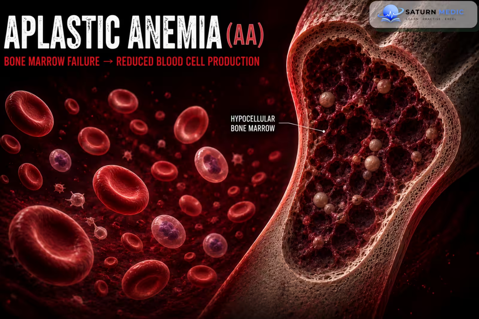
Aplastic Anemia (AA) is a rare, life-threatening bone marrow failure disorder characterized by pancytopenia and a markedly hypocellular bone marrow. It results from failure of hematopoietic stem cells to produce red blood cells, white blood cells, and platelets.
Also called
ICD-10
Specialty
Onset
Reviewed
-
OverviewOverview
-
Etiology & Risk FactorsEtiology & Risk Factors
-
PathophysiologyPathophysiology
-
Clinical PresentationClinical Presentation
-
History TakingHistory Taking
-
Physical ExaminationPhysical Examination
-
InvestigationsInvestigations
-
DiagnosisDiagnosis
-
ManagementManagement
-
ComplicationsComplications
-
PrognosisPrognosis
-
Key Points / Clinical PearlsKey Points / Clinical Pearls
Overview
Aplastic Anemia (AA) occurs when the bone marrow fails to produce adequate blood cells, leading to anemia, leukopenia, and thrombocytopenia (pancytopenia). Most acquired cases result from immune-mediated destruction of hematopoietic stem cells, although drugs, toxins, viral infections, radiation, and inherited disorders may also cause the disease.
Patients commonly present with fatigue, recurrent infections, and bleeding, while hepatosplenomegaly and lymphadenopathy are usually absent.
Etiology & Risk Factors
Idiopathic (Most Common) cause of Aplastic Anemia (AA)
No identifiable cause
Thought to be immune-mediated
Drug-Induced
Chloramphenicol
Carbamazepine
Phenytoin
Sulfonamides
Methimazole
Chemotherapy agents
Chemical Exposure
Benzene
Organic solvents
Pesticides
Radiation
Ionizing radiation
Radiotherapy
Viral Infections
Hepatitis viruses
Epstein-Barr virus (EBV)
Cytomegalovirus (CMV)
HIV
Parvovirus B19
Autoimmune Diseases
Systemic lupus erythematosus
Other autoimmune disorders
Inherited Disorders
Fanconi anemia
Dyskeratosis congenita
Shwachman-Diamond syndrome
-Risk Factors for Aplastic Anemia (AA)
Exposure to toxic chemicals
Cytotoxic drugs
Radiation exposure
Viral infections
Autoimmune disease
Family history of inherited bone marrow failure syndromes
Pathophysiology
Immune-mediated destruction or direct injury of hematopoietic stem cells → marked reduction in bone marrow stem cell population → hypocellular bone marrow with fatty replacement → decreased production of erythrocytes, leukocytes, and platelets → pancytopenia → anemia causes fatigue, neutropenia predisposes to infections, and thrombocytopenia leads to bleeding.
Clinical Presentation
-Symptoms of Aplastic Anemia (AA)
Fatigue
Weakness
Shortness of breath
Dizziness
Fever
Recurrent infections
Easy bruising
Gum bleeding
Epistaxis
Prolonged bleeding
Menorrhagia
-Signs of Aplastic Anemia (AA)
Pallor
Petechiae
Purpura
Ecchymoses
Fever (if infection is present)
-Usually absent in Aplastic Anemia (AA):
Lymphadenopathy
Hepatomegaly
Splenomegaly
History Taking
-Ask about:
- Fatigue
- Fever
- Recurrent infections
- Bleeding episodes
- Easy bruising
- Drug exposure
- Occupational chemical exposure
- Radiation exposure
- Recent viral illness
- Autoimmune disease
- Previous chemotherapy
Physical Examination
-General Examination
Look for:
Pallor
Fever
Petechiae
Purpura
Ecchymoses
-Systemic Examination
Assess for:
Signs of infection
Mucosal bleeding
-Confirm absence of:
Lymphadenopathy
Hepatomegaly
Splenomegaly
Investigations
Complete Blood Count (CBC)
Typical findings:
Low hemoglobin
Low white blood cell count
Low neutrophil count
Low platelet count
Pancytopenia
Low reticulocyte count
Peripheral Blood Film
May show:
Pancytopenia
Normocytic or mildly macrocytic red cells
No abnormal blast cells
Bone Marrow Aspiration and Biopsy (Gold Standard)
Characteristic findings:
Markedly hypocellular marrow
Fatty replacement
Marked reduction in hematopoietic cells
No malignant infiltration
Reticulocyte Count
Markedly reduced
Additional Laboratory Tests
Vitamin B12
Folate
Iron studies
Liver function tests
Renal function tests
Viral serology (HBV, HCV, HIV, EBV, CMV, Parvovirus B19)
Autoimmune screen
Flow Cytometry
Evaluate for paroxysmal nocturnal hemoglobinuria (PNH) clone
Cytogenetic Analysis
Exclude myelodysplastic syndrome and other marrow disorders
Diagnosis
Aplastic Anaemia · Classification by Cause
- Most common — CD8+ T-cells attack HSCs via IFN-γ and TNF-α
- Often follows viral illness (trigger unknown)
- Associated with HLA-DR2 alleles
- Responds to immunosuppression (ATG + CsA)
- Cytotoxic chemotherapy (busulfan, cyclophosphamide)
- Benzene (industrial)
- Chloramphenicol — classic cause
- NSAIDs (phenylbutazone, indomethacin)
- Carbamazepine, phenytoin
- Gold salts, sulfonamides, clozapine
- Seronegative hepatitis (non-A/B/C) — most common viral cause
- EBV, CMV
- HIV — direct HSC infection
- Parvovirus B19 — pure red cell aplasia (selective)
- Dengue fever — transient suppression
- Ionising radiation — dose-dependent direct HSC DNA damage
- Benzene — occupational (petroleum, rubber)
- Pesticides (organophosphates, DDT)
- Arsenic, toluene (glue sniffing)
- Rare — may remit after delivery
- Immune tolerance shift during pregnancy
- High maternal and foetal mortality if untreated
- HSCT deferred until post-partum if possible
- PNH — ~50% of AA have GPI-anchor deficient clone
- Eosinophilic fasciitis (Shulman syndrome)
- Thymoma (Good's syndrome)
- SLE — immune complex-mediated suppression
- Transfusion-associated GvHD
- Most common inherited AA — AR (FANCA mutation)
- DNA repair defect → HSC instability
- AA onset: childhood (median age 7–8 yrs)
- Café-au-lait spots, thumb/radial anomalies, short stature, microcephaly, renal anomalies
- ↑ Risk AML + solid tumours (HPV-related)
- Dx: DEB/MMC chromosomal breakage test
- Telomere biology disorder — DKC1, TERT, TERC, RTEL1
- Triad: nail dystrophy + oral leucoplakia + reticulate skin pigmentation
- AA in 80–90% by age 30
- Pulmonary fibrosis, liver disease, early greying
- Dx: lymphocyte telomere length (very short)
- Pure red cell aplasia; RPS19 mutation; steroid-responsive
- SBDS; neutropenia + exocrine pancreatic insufficiency
- ELANE/HAX1; neutropenia; G-CSF responsive
- AK2; absent myeloid + lymphoid; HSCT only
Aplastic Anaemia · Diagnostic Workup
Reticulocytes ↓↓ — inappropriately low (key)
Film: no dysplasia, no blasts
MCV may be ↑ (stress erythropoiesis)
No dysplasia = distinguishes from MDS
Hypocellularity <25% + fat cell replacement
No fibrosis, no infiltration, no dysplasia
Residual lymphocytes + plasma cells only
Viral serology — hepatitis A/B/C, EBV, CMV, HIV, Parvovirus B19
ANA, dsDNA — SLE
TFTs — hypothyroidism
LDH, uric acid — leukaemia/lymphoma
CT chest/abdomen — lymphadenopathy, thymoma
Present in ~50% of acquired AA
PNH clone >50% → eculizumab
Always test — changes management
NGS panel — DNMT3A, ASXL1, BCOR, PIGA
Telomere length — if DC suspected
DEB/MMC breakage test — if Fanconi suspected (age <40)
Unrelated donor search if no sibling match
Echo + PFTs — pre-transplant fitness
Ferritin — iron overload from transfusions
Minimise transfusions pre-HSCT
| Condition | Marrow Cellularity | Blasts | Dysplasia | Key distinguishing feature |
|---|---|---|---|---|
| Aplastic Anaemia | Hypocellular <25% | Absent | Absent | Fat replacement, empty marrow |
| MDS | Normo / hypercellular | <20% | Present | Dysplastic cells, cytogenetic abnormalities |
| AML | Hypercellular | ≥20% | Variable | Blast infiltration |
| B12 / Folate deficiency | Hypercellular | Absent | Megaloblastic | Macro-ovalocytes, hypersegmented neutrophils |
| PNH | Variable | Absent | Absent | CD55⁻/CD59⁻ on flow, haemolysis, thrombosis |
| Hypersplenism | Normal / ↑ | Absent | Absent | Massive splenomegaly |
Management
Supportive Care
Blood transfusions as required
Platelet transfusions
Prompt treatment of infections
Broad-spectrum antibiotics for febrile neutropenia
Good infection prevention measures
Immunosuppressive Therapy
For patients not suitable for transplantation:
Antithymocyte globulin (ATG)
Cyclosporine
Eltrombopag (in selected patients)
Hematopoietic Stem Cell Transplantation
First-line treatment for:
Severe Aplastic Anemia (AA)
Young patients
Suitable matched sibling donor available
Treat the Underlying Cause
Stop causative drugs
Avoid toxic chemical exposure
Treat associated infections
Manage autoimmune disease
Complications
- Severe bacterial infections
- Fungal infections
- Sepsis
- Major bleeding
- Intracranial hemorrhage
- Iron overload from repeated transfusions
- Relapse
- Evolution to myelodysplastic syndrome
- Acute myeloid leukemia
- Paroxysmal nocturnal hemoglobinuria (PNH)
Prognosis
The prognosis depends on disease severity, patient age, and response to treatment. With modern immunosuppressive therapy and hematopoietic stem cell transplantation, long-term survival has improved significantly. Severe untreated Aplastic Anemia (AA) has a high mortality due to infection and bleeding.
Key Points / Clinical Pearls
- Aplastic Anemia (AA) is a bone marrow failure syndrome causing pancytopenia.
- Most acquired cases are immune-mediated.
- Patients present with fatigue, infections, and bleeding.
- Lymphadenopathy and splenomegaly are usually absent.
- CBC shows pancytopenia with a low reticulocyte count.
- Bone marrow biopsy demonstrates a markedly hypocellular marrow.
- Stem cell transplantation is the preferred treatment for eligible patients with severe disease.
- Immunosuppressive therapy is the main alternative for non-transplant candidates.
- Prompt treatment of infections and bleeding is essential.
- Long-term follow-up is needed because of relapse and clonal evolution risks.
- Nimmana BK, Penney SW. National Center for Biotechnology Information (NIH). Aplastic Anemia, StatPearls.
- Killick SB, Cavenagh J, Ho A, et al. Guidelines for the Diagnosis and Management of Adult Aplastic Anaemia: A British Society for Haematology Guideline. Br J Haematol. 2024;204:784-804.
- MedlinePlus, National Library of Medicine (NIH). Aplastic Anemia: Medical Encyclopedia.
- National Center for Biotechnology Information (NIH). Hematopoietic Stem Cell Transplantation, StatPearls.
- National Center for Biotechnology Information (NIH). Fanconi Anemia, StatPearls.