

Clinical Subject Page
G6PD Deficiency
Glucose-6-Phosphate Dehydrogenase (G6PD) Deficiency is an X-linked inherited enzymatic disorder that predisposes red blood cells to oxidative damage, resulting in episodes of acute hemolytic anemia after exposure to oxidative stress
Also called
ICD-10
Specialty
Onset
Reviewed
-
OverviewOverview
-
Etiology & Risk FactorsEtiology & Risk Factors
-
PathophysiologyPathophysiology
-
Clinical PresentationClinical Presentation
-
History TakingHistory Taking
-
Physical ExaminationPhysical Examination
-
InvestigationsInvestigations
-
DiagnosisDiagnosis
-
ManagementManagement
-
ComplicationsComplications
-
PrognosisPrognosis
-
Key Points / Clinical PearlsKey Points / Clinical Pearls
Overview
Glucose-6-Phosphate Dehydrogenase (G6PD) Deficiency is the most common inherited red blood cell enzyme disorder worldwide. G6PD is essential for generating NADPH, which protects red blood cells from oxidative injury. Deficiency leads to episodic hemolysis triggered by infections, certain drugs, or fava beans. Most patients are asymptomatic between episodes
Etiology & Risk Factors
Glucose-6-Phosphate Dehydrogenase (G6PD) Deficiency is caused by mutations in the G6PD gene on the X chromosome, resulting in reduced G6PD enzyme activity.
Inheritance
- X-linked recessive
- Males are most commonly affected
- Females may be carriers or affected due to lyonization
G6PD Deficiency · Risk Factors & Triggers of Haemolysis
- Primaquine — most important; used in P. vivax/ovale
- Tafenoquine — newer; more potent trigger
- Chloroquine — mild trigger in severe deficiency
- Quinine — mild risk
- Dapsone — strong oxidant; high risk
- Nitrofurantoin — UTI treatment; avoid in G6PD
- Co-trimoxazole (trimethoprim-sulfamethoxazole)
- Nalidixic acid, ciprofloxacin (high doses)
- Chloramphenicol
- Aspirin (high dose) — low doses generally safe
- Phenacetin — withdrawn but still relevant historically
- Rasburicase — contraindicated (used in tumour lysis)
- Methylene blue — paradoxically worsens haemolysis in G6PD deficiency
- Vitamin K analogues (menadione)
- Fava beans (broad beans) — vicine + convicine → oxidative stress; causes "favism"
- Classic trigger in Mediterranean G6PD variant
- Haemolysis within hours of ingestion
- Can occur via breast milk (mother ate fava beans)
- Avoid all legumes in severe deficiency (Class I/II)
- Most common trigger overall — any acute infection
- Bacterial infections — pneumonia, typhoid, sepsis
- Viral — hepatitis A & B, CMV, EBV, influenza
- Malaria itself — oxidative stress from parasite
- Mechanism: activated neutrophils generate ROS → overwhelm deficient RBC defences
- Neonatal jaundice — most common presentation in neonates
- Appears day 2–3 of life (unlike physiological jaundice)
- Triggers: infection, maternal drug exposure (e.g. nitrofurantoin), naphthalene (mothballs)
- Risk of kernicterus if untreated
- Mandatory neonatal screening in endemic countries
- Naphthalene (mothballs) — common household trigger; especially dangerous in neonates
- Henna (mehndi) — contains lawsone; significant oxidant
- Methylene blue — worsens haemolysis (do not use as antidote)
- Industrial chemicals — nitro compounds, aniline dyes
- Diabetic ketoacidosis (DKA) — metabolic acidosis + oxidative stress
- Severe hypoglycaemia
- Strenuous exercise — oxidative burst
- Surgery / trauma — oxidative stress response
- Vitamin C in very high doses (IV) — pro-oxidant at high concentrations
- Paracetamol (acetaminophen) — safe at therapeutic doses
- Aspirin — safe at low doses (<1g/day)
- Chloroquine — generally safe for prophylaxis
- Penicillins, cephalosporins — safe
- Artemisinins — safe; preferred antimalarial in G6PD
- Proguanil — safe for malaria prophylaxis
Pathophysiology
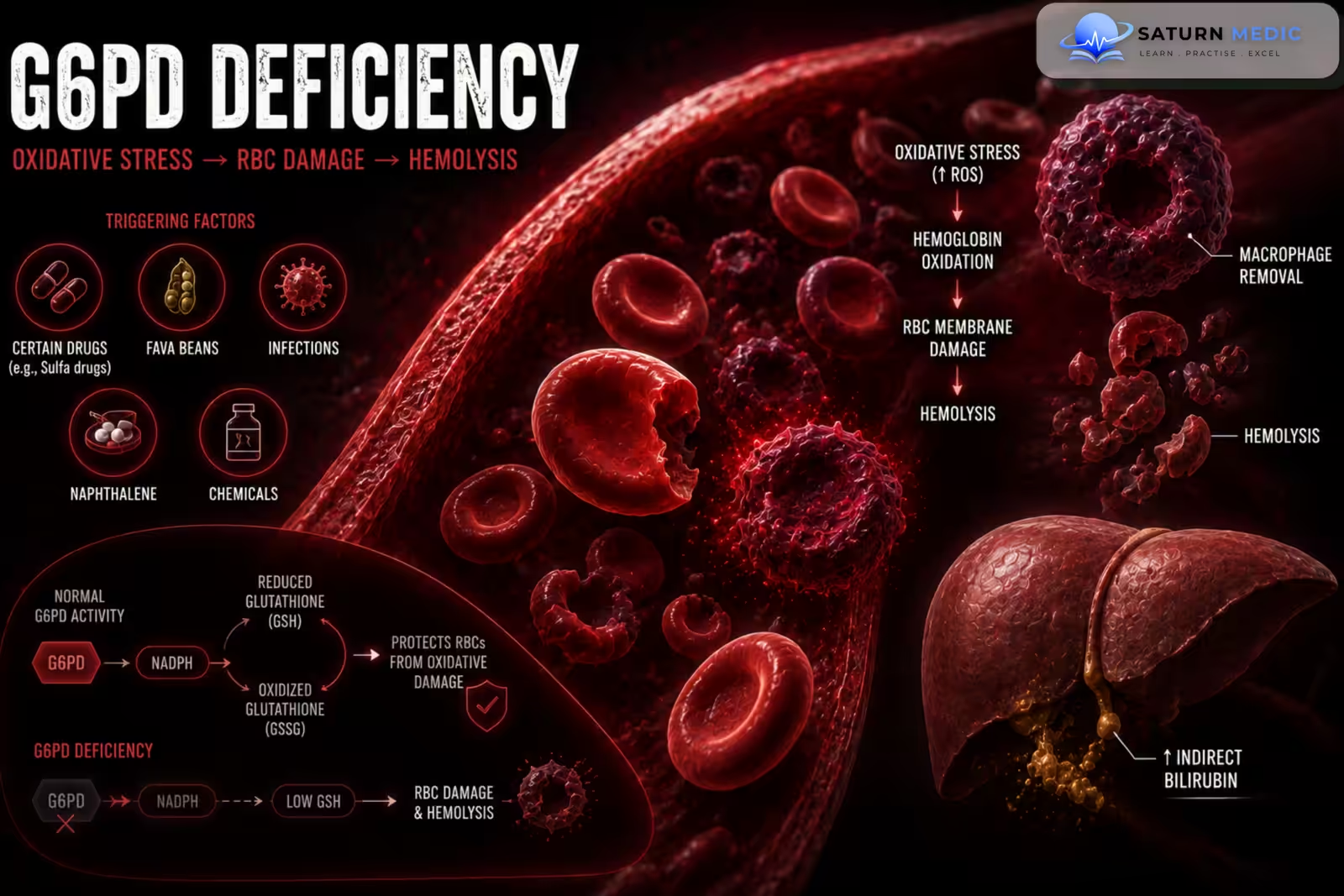
Inherited G6PD enzyme deficiency → reduced NADPH production → decreased regeneration of reduced glutathione → red blood cells become vulnerable to oxidative stress → oxidation and denaturation of hemoglobin → Heinz body formation → splenic macrophages remove Heinz bodies producing bite cells → intravascular and extravascular hemolysis → acute hemolytic anemia → increased bilirubin and reticulocytosis during recovery.
Clinical Presentation
Symptoms
-
Sudden fatigue
-
Weakness
-
Pallor
-
Jaundice
-
Dark urine
-
Back pain
-
Abdominal pain
-
Shortness of breath
Neonatal Presentation
-
Neonatal jaundice
-
Severe hyperbilirubinemia
-
Kernicterus (rare but serious)
Signs
-
Pallor
-
Jaundice
-
Tachycardia
-
Mild splenomegaly (occasionally during hemolytic episodes)
History Taking
-Ask about:
- Recent infection
- New medications
- Fava bean ingestion
- Dark urine
- Jaundice
- Previous hemolytic episodes
- Neonatal jaundice
- Family history
- Ethnic background
- Blood transfusions
Physical Examination
-General Examination
Look for:
-
Pallor
-
Jaundice
-
Tachycardia
-
Fever if infection is present
-Systemic Examination
Assess for:
-
Mild splenomegaly
-
Signs of dehydration
-
Evidence of the triggering illness
Investigations
-Complete Blood Count (CBC)
Typical findings during hemolysis:
-
Low hemoglobin
-
Increased reticulocyte count (after marrow response)
-Peripheral Blood Film
Characteristic findings:
-
Bite cells
-
Blister cells
-
Polychromasia
-
Heinz bodies (seen with supravital stain)
-Hemolysis Screen
-
Increased indirect bilirubin
-
Increased LDH
-
Low haptoglobin
-
Increased reticulocyte count
G6PD Enzyme Assay (Diagnostic Test)
-
Confirms G6PD deficiency.
-
May be falsely normal during an acute hemolytic episode due to the presence of young red blood cells with higher enzyme activity.
-
Repeat testing several weeks after recovery if clinical suspicion remains high.
Additional Investigations
-
Urinalysis (hemoglobinuria)
-
Renal function tests
-
Direct antiglobulin (Coombs) test to exclude autoimmune hemolysis if indicated
Diagnosis
Diagnosis is based on:
- Typical history of episodic hemolysis after oxidative stress
- Hemolysis laboratory findings
- Characteristic peripheral blood film
- Reduced G6PD enzyme activity on enzyme assay
Management
Acute Hemolytic Episode
-
Immediately stop the triggering drug or exposure
-
Treat underlying infection
-
Maintain adequate hydration
-
Monitor renal function
-
Folic acid supplementation if appropriate
-
Blood transfusion for severe symptomatic anemia
Long-Term Management
-
Avoid oxidant drugs
-
Avoid fava beans
-
Prompt treatment of infections
-
Patient education regarding triggers
-
Genetic counseling when appropriate
Neonatal Management
-
Phototherapy
-
Exchange transfusion if severe hyperbilirubinemia develops
Complications
- Acute hemolytic anemia
- Severe jaundice
- Acute kidney injury
- Hemoglobinuria
- Neonatal kernicterus
- Rarely, life-threatening anemia
Prognosis
The prognosis is excellent for most patients when oxidative triggers are avoided. Hemolytic episodes are usually self-limited, and red blood cell production recovers once the trigger is removed. Long-term survival is normal with appropriate education and prevention.
Key Points / Clinical Pearls
- G6PD Deficiency is an X-linked inherited enzyme disorder.
- It is the most common inherited red blood cell enzyme deficiency worldwide.
- Hemolysis is triggered by oxidative stress.
- Common triggers include infections, oxidant drugs, and fava beans.
- Peripheral blood film shows bite cells and Heinz bodies (with supravital stain).
- G6PD enzyme assay confirms the diagnosis but may be falsely normal during acute hemolysis.
- Stop the trigger immediately and provide supportive care.
- Blood transfusion is reserved for severe hemolysis.
- Avoidance of triggers is the cornerstone of long-term management.
- Prognosis is excellent with proper education and trigger avoidance.
- Mak GK. National Center for Biotechnology Information (NIH). Glucose-6-Phosphate Dehydrogenase Deficiency, StatPearls.
- Luzzatto L, Ally M, Notaro R. Glucose-6-Phosphate Dehydrogenase Deficiency. Blood. 2020;136:1225-1240. ASH Blood Journal.
- Sawaftah MA, Babi J, Abuzayda HA, Ahmed G. Two Cases of Methemoglobinemia Secondary to Favism in Pediatric Patients With Unknown G6PD Deficiency. Cureus. 2026. PMC12888067.
- Nannelli C, Bosman A, Cunningham J, Dugue PA, Luzzatto L. Genetic Variants Causing G6PD Deficiency: Clinical and Biochemical Data Support New WHO Classification. Br J Haematol. 2023;202:1024-1032.
- MedlinePlus, National Library of Medicine (NIH). G6PD Deficiency: Genetics Home Reference.