

Clinical Subject Page
Autoimmune Hemolytic Anemia (AIHA)
Autoimmune Hemolytic Anemia (AIHA) is an acquired immune-mediated disorder in which autoantibodies target red blood cells, causing their premature destruction and resulting in hemolytic anemia. Hemolysis may occur within the spleen (extravascular) or the circulation (intravascular)
Also called
ICD-10
Specialty
Onset
Reviewed
-
OverviewOverview
-
Etiology & Risk FactorsEtiology & Risk Factors
-
PathophysiologyPathophysiology
-
Clinical PresentationClinical Presentation
-
History TakingHistory Taking
-
Physical ExaminationPhysical Examination
-
InvestigationsInvestigations
-
DiagnosisDiagnosis
-
ManagementManagement
-
ComplicationsComplications
-
PrognosisPrognosis
-
Key Points / Clinical PearlsKey Points / Clinical Pearls
Overview
Autoimmune Hemolytic Anemia (AIHA) occurs when the immune system produces antibodies against red blood cells. Warm AIHA (IgG-mediated) is the most common type and usually causes extravascular hemolysis in the spleen, whereas Cold AIHA (IgM-mediated) activates complement and often causes hemolysis after cold exposure.
Patients typically present with anemia, jaundice, reticulocytosis, and laboratory evidence of hemolysis. The Direct Antiglobulin (Coombs) Test is the key diagnostic investigation.
Etiology & Risk Factors
-Primary (Idiopathic) cause of Autoimmune Hemolytic Anemia (AIHA)
No identifiable underlying cause
-Secondary Causes of Autoimmune Hemolytic Anemia (AIHA)
Autoimmune Diseases
Systemic lupus erythematosus
Rheumatoid arthritis
Autoimmune thyroid disease
Lymphoproliferative Disorders
Chronic lymphocytic leukemia (CLL)
Non-Hodgkin lymphoma
Hodgkin lymphoma
Infections
Mycoplasma pneumoniae
Epstein-Barr virus (EBV)
Cytomegalovirus (CMV)
HIV
Drugs
Penicillin
Cephalosporins
Methyldopa
Fludarabine
-Risk Factors
Autoimmune disease
Lymphoid malignancy
Recent infection
Certain medications
Older age (especially Warm AIHA)
Pathophysiology
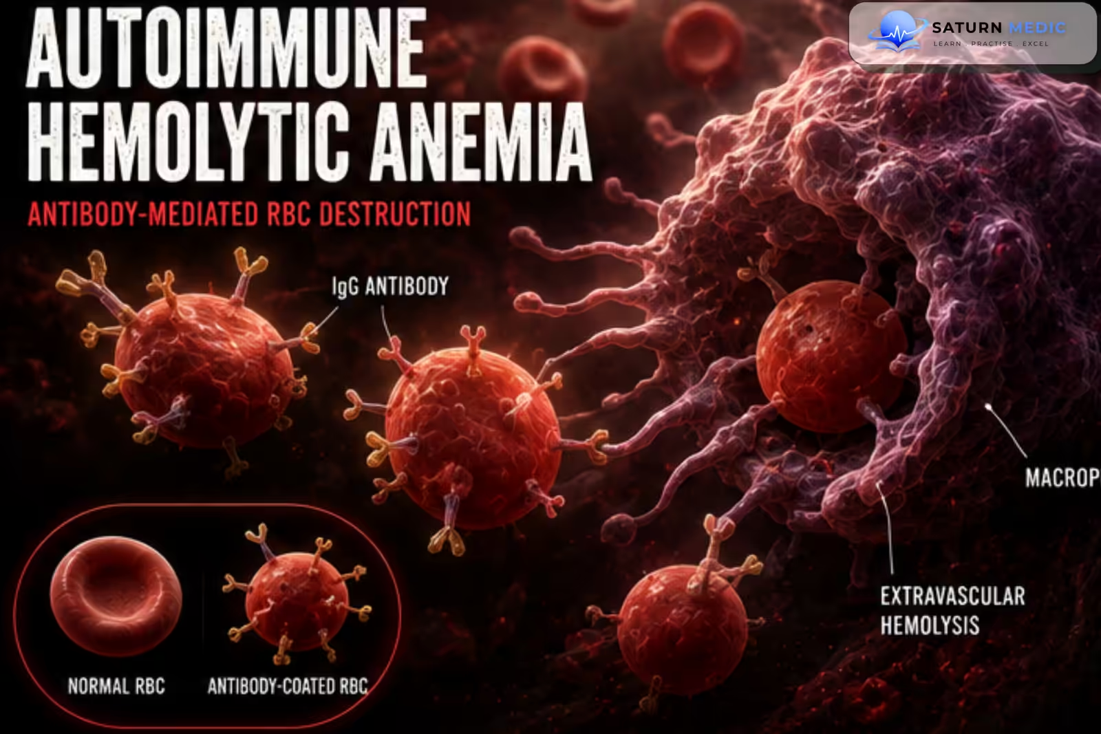
Autoantibody formation against red blood cell surface antigens → antibody-coated red blood cells are recognized by macrophages or activate complement → premature red blood cell destruction → hemolytic anemia.
-Warm AIHA
IgG antibodies bind red blood cells at body temperature → splenic macrophages remove antibody-coated RBCs → extravascular hemolysis.
-Cold AIHA
IgM antibodies bind RBCs at low temperatures → complement activation → intravascular and extravascular hemolysis, primarily in the liver.
Clinical Presentation
-Symptoms
Fatigue
Weakness
Pallor
Jaundice
Dark urine
Shortness of breath
Dizziness
Palpitations
-Signs
Pallor
Jaundice
Splenomegaly (especially Warm AIHA)
Mild hepatomegaly
Tachycardia
-Cold Autoimmune Hemolytic Anemia (AIHA) Features
Acrocyanosis
Raynaud-like symptoms
Hemolysis after cold exposure
History Taking
-Ask about:
- Fatigue
- Jaundice
- Dark urine
- Previous hemolytic episodes
- Cold sensitivity
- Recent infections
- Autoimmune disease
- Malignancy
- Medication history
- Blood transfusions
- Family history
Physical Examination
-General Examination
Look for:
Pallor
Jaundice
Fever
Tachycardia
-Abdominal Examination
Assess for:
Splenomegaly
Hepatomegaly
-Peripheral Examination
Look for:
Acrocyanosis
Peripheral cyanosis
Signs of underlying autoimmune disease
Investigations
-Complete Blood Count (CBC)
Typical findings:
Low hemoglobin
Increased reticulocyte count
Variable MCV (may be increased due to reticulocytosis)
-Peripheral Blood Film
May show:
Spherocytes (Warm AIHA)
Polychromasia
RBC agglutination (Cold AIHA)
-Hemolysis Screen
Increased indirect bilirubin
Increased LDH
Low haptoglobin
Increased reticulocyte count
-Direct Antiglobulin (Coombs) Test (Gold Standard)
Positive in most cases
Detects IgG and/or complement bound to red blood cells
Indirect Antiglobulin Test
May identify circulating antibodies
Additional Investigations
To identify underlying causes:
ANA
Serum protein electrophoresis
Immunoglobulin levels
Viral serology (HIV, EBV, CMV, Mycoplasma)
CT imaging if lymphoma is suspected
Bone marrow examination when indicated
Diagnosis
Diagnosis is based on:
- Clinical features of hemolysis
- Positive Direct Antiglobulin (Coombs) Test
- Laboratory evidence of hemolysis
- Identification of any underlying disease
Management
-Initial Management
Treat the underlying cause
Supportive care
Folic acid supplementation
Blood transfusion if severe symptomatic anemia (when necessary)
-Warm AIHA
First-line:
Corticosteroids (Prednisolone)
Second-line:
Rituximab
Splenectomy (selected patients)
Immunosuppressive agents (e.g., azathioprine, mycophenolate mofetil, cyclophosphamide)
-Cold AIHA
Avoid cold exposure
Rituximab is first-line therapy for symptomatic disease
Complement inhibitors (selected patients)
Corticosteroids are generally less effective than in Warm AIHA
Complications
- Severe anemia
- Heart failure
- Thromboembolism
- Hemolytic crisis
- Gallstones
- Splenomegaly
- Treatment-related immunosuppression
- Relapse
Prognosis
The prognosis depends on the underlying cause and the response to treatment. Many patients with Warm AIHA respond well to corticosteroids, although relapses are common. Cold AIHA tends to follow a chronic course but can often be controlled with avoidance of cold exposure and targeted therapy. Early diagnosis and treatment improve outcomes.
Key Points / Clinical Pearls
- Autoimmune Hemolytic Anemia (AIHA) is an acquired immune-mediated hemolytic anemia.
- The two major types are Warm AIHA (IgG) and Cold AIHA (IgM).
- Warm AIHA is the most common form.
- Patients present with anemia, jaundice, and laboratory evidence of hemolysis.
- The Direct Antiglobulin (Coombs) Test is the key diagnostic test.
- Warm AIHA commonly shows spherocytes on the peripheral blood film.
- Corticosteroids are the first-line treatment for Warm AIHA.
- Rituximab is an important therapy for refractory Warm AIHA and symptomatic Cold AIHA.
- Always investigate for underlying autoimmune disease, infection, or lymphoproliferative malignancy.
- Prognosis is generally good with timely diagnosis and appropriate treatment.
- Tripathi AK, Chuda R. National Center for Biotechnology Information (NIH). Laboratory Evaluation of Immune Hemolytic Anemias, StatPearls.
- Jager U, Barcellini W, Broome CM, et al. Diagnosis and Treatment of Autoimmune Hemolytic Anemia in Adults: Recommendations From the First International Consensus Meeting. Blood Rev. 2020;41:100648.
- Autoimmune Hemolytic Anemia in the Pediatric Setting. J Clin Med. 2021;10:216. PMC7828053.
- Barcellini W, Fattizzo B. Autoimmune Hemolytic Anemia: Current Understanding of Pathogenesis and Treatment. Front Immunol. EHA Library.
- MedlinePlus, National Library of Medicine (NIH). Autoimmune Hemolytic Anemia: Medical Encyclopedia.