

Clinical Subject Page
Polycythemia Vera (PV)
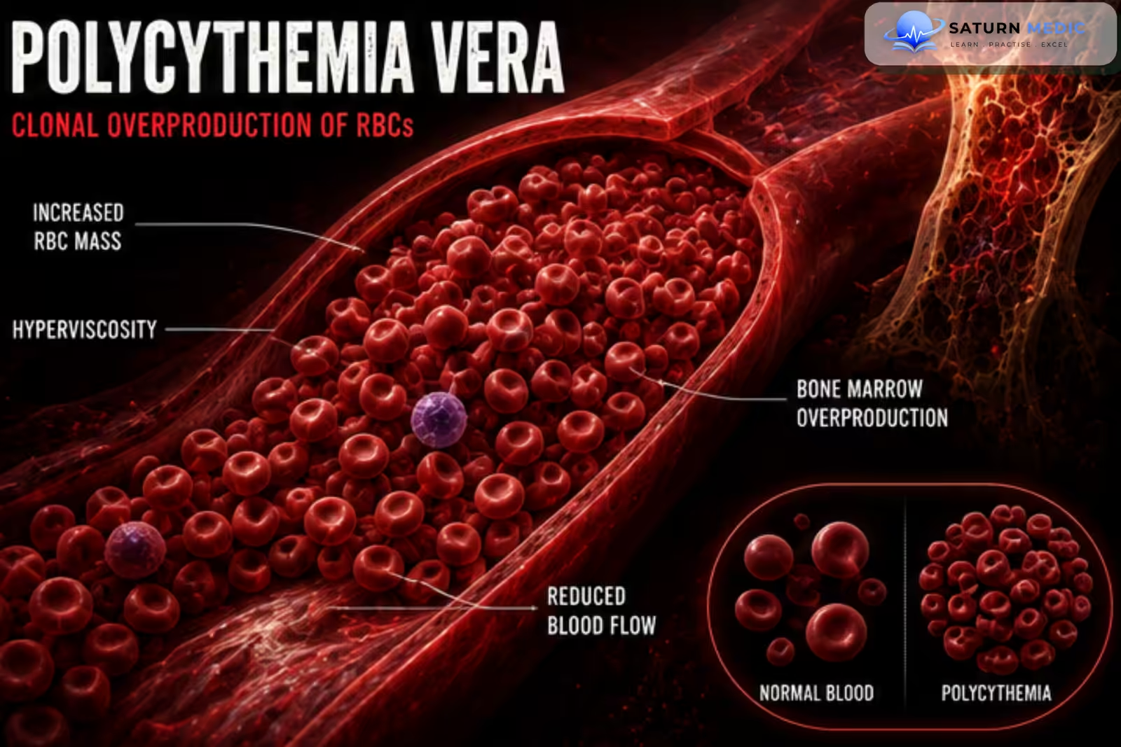
Polycythemia Vera (PV) is a chronic myeloproliferative neoplasm (MPN) characterized by clonal overproduction of red blood cells, often accompanied by increased white blood cells and platelets. Most cases are associated with a JAK2 mutation.
Also called
ICD-10
Specialty
Onset
Reviewed
-
OverviewOverview
-
Etiology & Risk FactorsEtiology & Risk Factors
-
PathophysiologyPathophysiology
-
Clinical PresentationClinical Presentation
-
History TakingHistory Taking
-
Physical ExaminationPhysical Examination
-
InvestigationsInvestigations
-
DiagnosisDiagnosis
-
ManagementManagement
-
ComplicationsComplications
-
PrognosisPrognosis
-
Key Points / Clinical PearlsKey Points / Clinical Pearls
Overview
Polycythemia Vera (PV) is a myeloproliferative neoplasm caused by constitutive activation of the JAK-STAT signaling pathway, usually due to a JAK2 mutation. Excess production of red blood cells increases blood viscosity, leading to thrombosis, while abnormal platelet function may paradoxically increase bleeding risk.
Without treatment, PV may progress to myelofibrosis or acute myeloid leukemia (AML).
Etiology & Risk Factors
Polycythemia Vera (PV) is caused by an acquired mutation in a hematopoietic stem cell.
Common Genetic Mutations
- JAK2 V617F mutation (≈95% of cases)
- JAK2 exon 12 mutation (most remaining cases)
-Risk Factors
- Increasing age
- Male sex
- Family history (rare)
- Previous exposure to ionizing radiation (possible association)
Pathophysiology
JAK2 mutation → constitutive activation of the JAK-STAT pathway → erythropoietin-independent proliferation of hematopoietic stem cells → excessive production of red blood cells, often with leukocytosis and thrombocytosis → increased blood viscosity → impaired microcirculation and hypercoagulability → increased risk of arterial and venous thrombosis → progressive bone marrow fibrosis or transformation to acute leukemia in some patients.
Clinical Presentation
-Symptoms
-
Headache
-
Dizziness
-
Fatigue
-
Blurred vision
-
Tinnitus
-
Pruritus (especially after a warm bath)
-
Burning pain and redness of the hands or feet (erythromelalgia)
-
Night sweats
-
Weight loss
-Signs
-
Plethoric (ruddy) complexion
-
Hypertension
-
Splenomegaly
-
Hepatomegaly (less common)
-
Thrombotic events
-Thrombotic Complications
-
Stroke
-
Transient ischemic attack
-
Myocardial infarction
-
Deep vein thrombosis
-
Pulmonary embolism
-
Budd-Chiari syndrome
-
Portal vein thrombosis
-Bleeding Manifestations
-
Epistaxis
-
Gingival bleeding
-
Easy bruising
-
Gastrointestinal bleeding
History Taking
-Ask about:
- Headache
- Dizziness
- Visual disturbance
- Pruritus
- Erythromelalgia
- Previous thrombosis
- Bleeding episodes
- Smoking history
- Symptoms of sleep apnea
- Weight loss
- Night sweats
- Family history
- Cardiovascular risk factors
Physical Examination
-General Examination
Look for:
-
Plethoric facial appearance
-
Hypertension
-
Cyanosis
-
Digital ischemia
-Abdominal Examination
Assess for:
-
Splenomegaly
-
Hepatomegaly
-Peripheral Examination
Look for:
-
-
Erythromelalgia
-
Signs of thrombosis
-
Signs of bleeding
-
Investigations
-Complete Blood Count (CBC)
Typical findings:
-
Increased hemoglobin
-
Increased hematocrit
-
Increased RBC count
-
Leukocytosis (common)
-
Thrombocytosis (common)
-Peripheral Blood Film
May show:
-
Increased red blood cells
-
Thrombocytosis
-
Neutrophilia
-Serum Erythropoietin (EPO)
-
Low (characteristic finding)
-JAK2 Mutation Testing
-
JAK2 V617F mutation (positive in ~95%)
-
JAK2 exon 12 mutation if V617F is negative
-Bone Marrow Biopsy
Typical findings:
-
Hypercellular marrow
-
Panmyelosis
-
Increased erythroid, granulocytic, and megakaryocytic proliferation
-Additional Investigations
-
-
Serum uric acid (often elevated)
-
LDH
-
Iron studies
-
Renal function tests
-
Liver function tests
-
Oxygen saturation (to exclude secondary polycythemia)
-
Abdominal ultrasound (splenomegaly)
-
Diagnosis
Diagnosis is based on:
- Elevated hemoglobin or hematocrit (or increased red cell mass)
- Bone marrow panmyelosis
- Presence of a JAK2 mutation
- Low serum erythropoietin level (minor criterion)\
Management
-Low-Risk Patients
-
Regular phlebotomy (venesection) to maintain hematocrit <45%
-
Low-dose aspirin (unless contraindicated)
-
Control cardiovascular risk factors
-High-Risk Patients
High risk includes:
-
Age ≥60 years
-
Previous thrombosis
Treatment:
-
Phlebotomy
-
Low-dose aspirin
-
Cytoreductive therapy
Cytoreductive Therapy
First-line:
-
Hydroxyurea
Alternative options:
-
Pegylated interferon-α
-
Ruxolitinib (particularly for hydroxyurea intolerance or resistance)
Supportive Management
-
Smoking cessation
-
Adequate hydration
-
Control hypertension, diabetes, and hyperlipidemia
-
Treat pruritus if present
Complications
- Arterial thrombosis
- Venous thrombosis
- Budd-Chiari syndrome
- Stroke
- Myocardial infarction
- Pulmonary embolism
- Major bleeding
- Hyperuricemia
- Gout
- Myelofibrosis
- Acute myeloid leukemia (AML)
Prognosis
With appropriate treatment, many patients have a near-normal life expectancy. Thrombosis remains the leading cause of morbidity and mortality. Long-term follow-up is essential because some patients may develop post-polycythemic myelofibrosis or transform to acute myeloid leukemia.
Key Points / Clinical Pearls
- Polycythemia Vera (PV) is a chronic myeloproliferative neoplasm.
- JAK2 V617F mutation is present in about 95% of patients.
- Serum erythropoietin (EPO) is typically low.
- Patients commonly present with headache, dizziness, and aquagenic pruritus.
- Increased blood viscosity predisposes to arterial and venous thrombosis.
- CBC usually shows erythrocytosis with leukocytosis and thrombocytosis.
- Phlebotomy and low-dose aspirin are the main initial treatments.
- Hydroxyurea is first-line cytoreductive therapy for high-risk patients.
- Maintain hematocrit <45% to reduce thrombotic risk.
- Long-term complications include myelofibrosis and acute myeloid leukemia (AML).
- National Center for Biotechnology Information (NIH). Polycythemia, StatPearls.
- Tefferi A, Barbui T. Polycythemia Vera: 2024 Update on Diagnosis, Risk-Stratification, and Management. Am J Hematol. 2023;98:1465-1487. DOI: 10.1002/ajh.27002.
- Khoury JD, Solary E, Abla O, et al. The 5th Edition of the World Health Organization Classification of Haematolymphoid Tumours: Myeloid and Histiocytic/Dendritic Neoplasms. Leukemia. 2022;36:1703-1719.
- MedlinePlus, National Library of Medicine (NIH). Polycythemia Vera: Medical Encyclopedia.
- National Center for Biotechnology Information (NIH). Myeloproliferative Disease, StatPearls.