

Clinical Subject Page
Acute Myeloid Leukemia (AML)
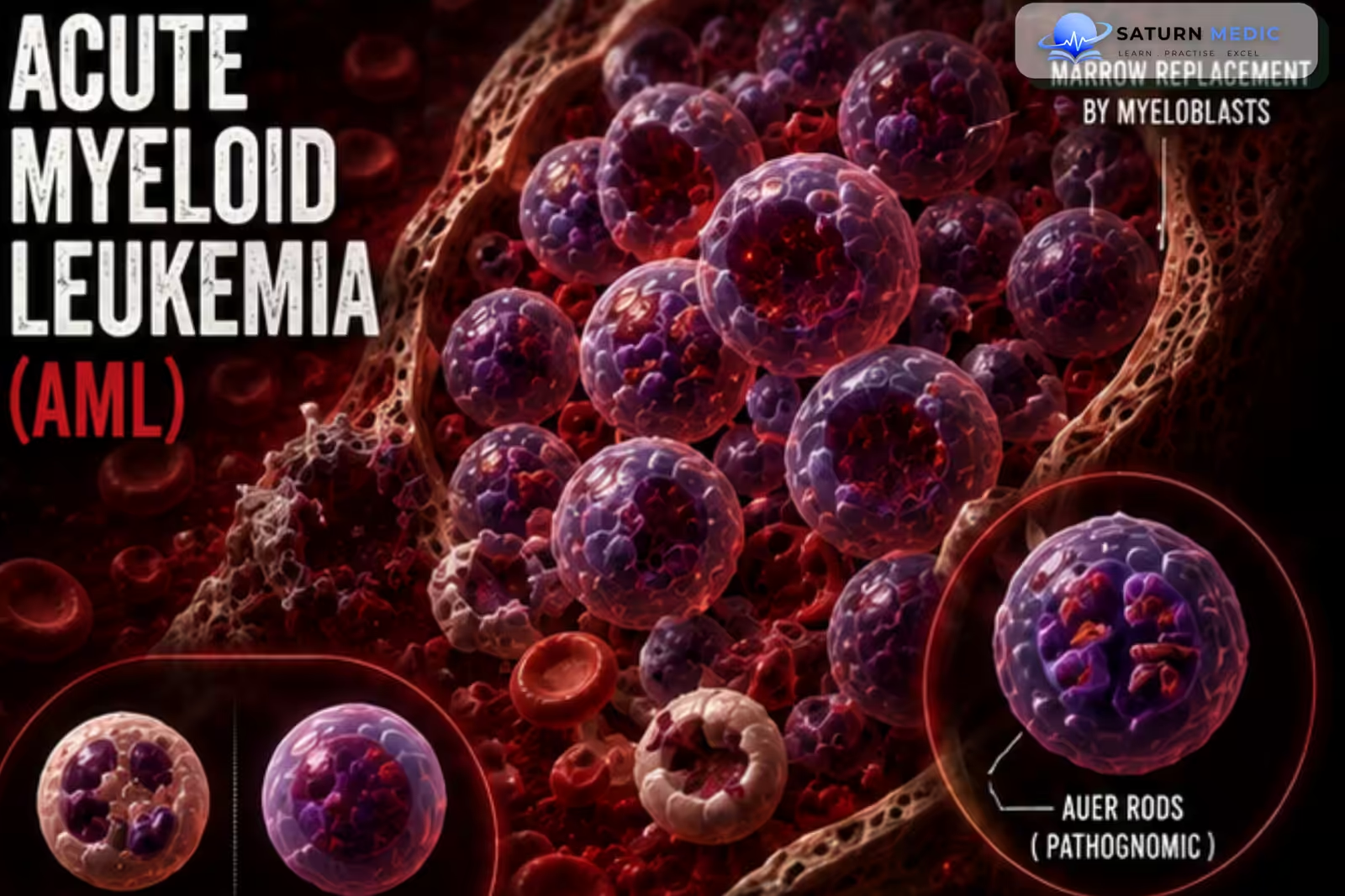
Acute Myeloid Leukemia (AML) is an aggressive hematologic malignancy caused by uncontrolled proliferation of abnormal immature myeloid precursor cells (myeloblasts) in the bone marrow. The abnormal cells suppress normal blood-cell production and may infiltrate other tissues
Also called
ICD-10
Specialty
Onset
Reviewed
-
OverviewOverview
-
Etiology & Risk FactorsEtiology & Risk Factors
-
PathophysiologyPathophysiology
-
Clinical PresentationClinical Presentation
-
History TakingHistory Taking
-
Physical ExaminationPhysical Examination
-
InvestigationsInvestigations
-
DiagnosisDiagnosis
-
ManagementManagement
-
ComplicationsComplications
-
PrognosisPrognosis
-
Key Points / Clinical PearlsKey Points / Clinical Pearls
Overview
Acute Myeloid Leukemia (AML) develops from malignant transformation of myeloid precursor cells, causing rapid accumulation of blasts in the bone marrow. This leads to anemia, neutropenia, and thrombocytopenia, producing fatigue, infections, and bleeding. Acute Myeloid Leukemia (AML) can occur at any age but is particularly common in older adults
Etiology & Risk Factors
The exact cause is often unknown. Acute Myeloid Leukemia (AML) results from acquired genetic and molecular abnormalities that cause:
- Uncontrolled proliferation
- Impaired differentiation
- Accumulation of immature myeloid blasts
-Risk Factors
- Increasing age
- Previous chemotherapy
- Previous radiotherapy
- Benzene exposure
- Smoking
- Myelodysplastic Syndrome (MDS)
- Myeloproliferative neoplasms
- Previous aplastic anemia
- Down syndrome and other genetic disorders
- Previous treatment with topoisomerase II inhibitors
Pathophysiology
Genetic mutations in a hematopoietic stem/progenitor cell → abnormal myeloid precursor proliferation and impaired differentiation → accumulation of myeloblasts in the bone marrow → suppression of normal hematopoiesis → anemia, neutropenia, and thrombocytopenia → infiltration of peripheral tissues such as skin, gums, spleen, and CNS in some patients.
Clinical Presentation
Symptoms
-
Fatigue
-
Weakness
-
Pallor
-
Fever
-
Recurrent infections
-
Easy bruising
-
Bleeding
-
Bone pain
-
Weight loss
-
Shortness of breath
Signs
-
Pallor
-
Fever
-
Petechiae
-
Ecchymoses
-
Lymphadenopathy
-
Hepatosplenomegaly
-
Gingival hypertrophy
-
Skin infiltration
-
Bone tenderness
History Taking
-Ask about:
- Fatigue
- Fever
- Recurrent infections
- Bleeding
- Easy bruising
- Bone pain
- Weight loss
- Night sweats
- Shortness of breath
- Previous chemotherapy or radiotherapy
- Occupational exposure to benzene
- Previous MDS or other hematologic disorders
- Family history
- Neurological symptoms
Physical Examination
-General Examination
Look for:
-
Pallor
-
Fever
-
Weight loss
-
Petechiae
-
Ecchymoses
-
Signs of infection
-Lymph Node Examination
Assess for:
-
Cervical
-
Axillary
-
Inguinal lymphadenopathy
-Abdominal Examination
Look for:
-
Hepatomegaly
-
Splenomegaly
-Oral Examination
Look for:
-
Gingival hypertrophy
-
Mucosal bleeding
-
Oral infections
-Skin Examination
Assess for:
-
Petechiae
-
Ecchymoses
-
Leukemic skin infiltration
Investigations
Complete Blood Count (CBC)
May show:
-
Anemia
-
Thrombocytopenia
-
Neutropenia
-
Leukocytosis, normal WBC, or leukopenia
-
Circulating blasts
Peripheral Blood Film
May show:
-
Myeloblasts
-
Auer rods
-
Anemia
-
Thrombocytopenia
-
Abnormal WBC count
Important Finding
Auer rods strongly support myeloid differentiation.
Bone Marrow Aspiration & Biopsy — Gold Standard
Typical findings:
-
Increased myeloblasts
-
Usually ≥20% blasts in the bone marrow or peripheral blood
Some genetically defined AML entities can be diagnosed without meeting the 20% blast threshold.
Flow Cytometry
Used to establish myeloid lineage.
Common markers include:
-
CD13
-
CD33
-
CD117
-
MPO
-
HLA-DR
-
CD34 in many cases
Cytogenetic & Molecular Testing
Important abnormalities include:
-
t(8;21) / RUNX1-RUNX1T1
-
inv(16) / CBFB-MYH11
-
PML-RARA
-
FLT3 mutations
-
NPM1 mutations
-
CEBPA mutations
-
TP53 abnormalities
These findings are essential for classification, prognosis, and treatment selection.
Coagulation Studies
Especially important when Acute Promyelocytic Leukemia (APL) is suspected:
-
PT
-
aPTT
-
Fibrinogen
-
D-dimer
Additional Investigations
-
Urea and electrolytes
-
Creatinine
-
Liver function tests
-
LDH
-
Uric acid
-
Blood cultures if febrile
-
Chest imaging if infection suspected
-
Baseline cardiac assessment before anthracyclines
Diagnosis
Diagnosis is based on:
- CBC and peripheral blood film
- Bone marrow examination
- Blast percentage
- Flow cytometry
- Cytogenetic and molecular studies
Classification identifies the specific genetic and molecular subtype of Acute Myeloid Leukemia (AML).
Management
-Initial Management
Treatment depends on:
-
Age
-
Fitness
-
Cytogenetic/molecular risk
-
Comorbidities
-
AML subtype
Fit Patients
-Intensive induction chemotherapy may include:
-
Cytarabine
-
An anthracycline such as daunorubicin or idarubicin
This is commonly known as “7+3” induction.
Targeted agents may be added depending on molecular abnormalities.
-Consolidation
After achieving remission:
-
High-dose cytarabine in selected patients
-
Allogeneic hematopoietic stem cell transplantation for appropriate intermediate/high-risk patients
-Older / Unfit Patients
Common approaches include:
-
Azacitidine + venetoclax
-
Other lower-intensity or targeted regimens depending on molecular findings and patient fitness
-Acute Promyelocytic Leukemia (APL)
APL is a medical emergency because of severe coagulopathy.
Treatment commonly involves:
-
All-trans retinoic acid (ATRA)
-
Arsenic trioxide
-
± chemotherapy depending on risk
ATRA should be started immediately when APL is strongly suspected.
Supportive Care
-
Red blood cell transfusions
-
Platelet transfusions
-
Prompt treatment of infections
-
Tumor lysis syndrome prevention
-
Antimicrobial prophylaxis when appropriate
-
Management of chemotherapy complications
Complications
- Severe anemia
- Severe infections
- Bleeding
- Disseminated intravascular coagulation, especially in APL
- Tumor lysis syndrome
- Leukostasis
- CNS involvement
- Organ infiltration
- Treatment-related complications
- Relapse
Prognosis
Prognosis varies considerably according to age, overall fitness, cytogenetic abnormalities, molecular mutations, treatment response, and measurable residual disease (MRD). Some genetically favorable forms have excellent outcomes, while adverse-risk Acute Myeloid Leukemia (AML) may require stem cell transplantation and has a higher relapse risk.
Key Points / Clinical Pearls
- Acute Myeloid Leukemia (AML) is a malignancy of immature myeloid cells.
- It is more common in older adults.
- Common presentation: anemia + infection + bleeding.
- Peripheral blood may show myeloblasts and Auer rods.
- Bone marrow examination is essential for diagnosis.
- Flow cytometry establishes myeloid lineage.
- Cytogenetic and molecular testing guides prognosis and treatment.
- PML-RARA identifies Acute Promyelocytic Leukemia (APL).
- Suspected APL requires immediate ATRA.
- Fit patients may receive intensive induction such as 7+3.
- Azacitidine + venetoclax is commonly used in many older/unfit patients.
- Allogeneic stem cell transplantation is used in selected patients at significant risk of relapse.
- Vakiti A, Reynolds SB, Mewawalla P. National Center for Biotechnology Information (NIH). Acute Myeloid Leukemia, StatPearls.
- Döhner H, Wei AH, Appelbaum FR, et al. Diagnosis and Management of AML in Adults: 2022 Recommendations From an International Expert Panel on Behalf of the ELN. Blood. 2022;140:1345-1377.
- National Comprehensive Cancer Network (NCCN). NCCN Clinical Practice Guidelines in Oncology: Acute Myeloid Leukemia.
- National Cancer Institute (NIH). Adult Acute Myeloid Leukemia Treatment (PDQ), Health Professional Version.
- MedlinePlus, National Library of Medicine (NIH). Acute Myelocytic Leukemia: Medical Encyclopedia.