

Clinical Subject Page
Hereditary Spherocytosis (HS)
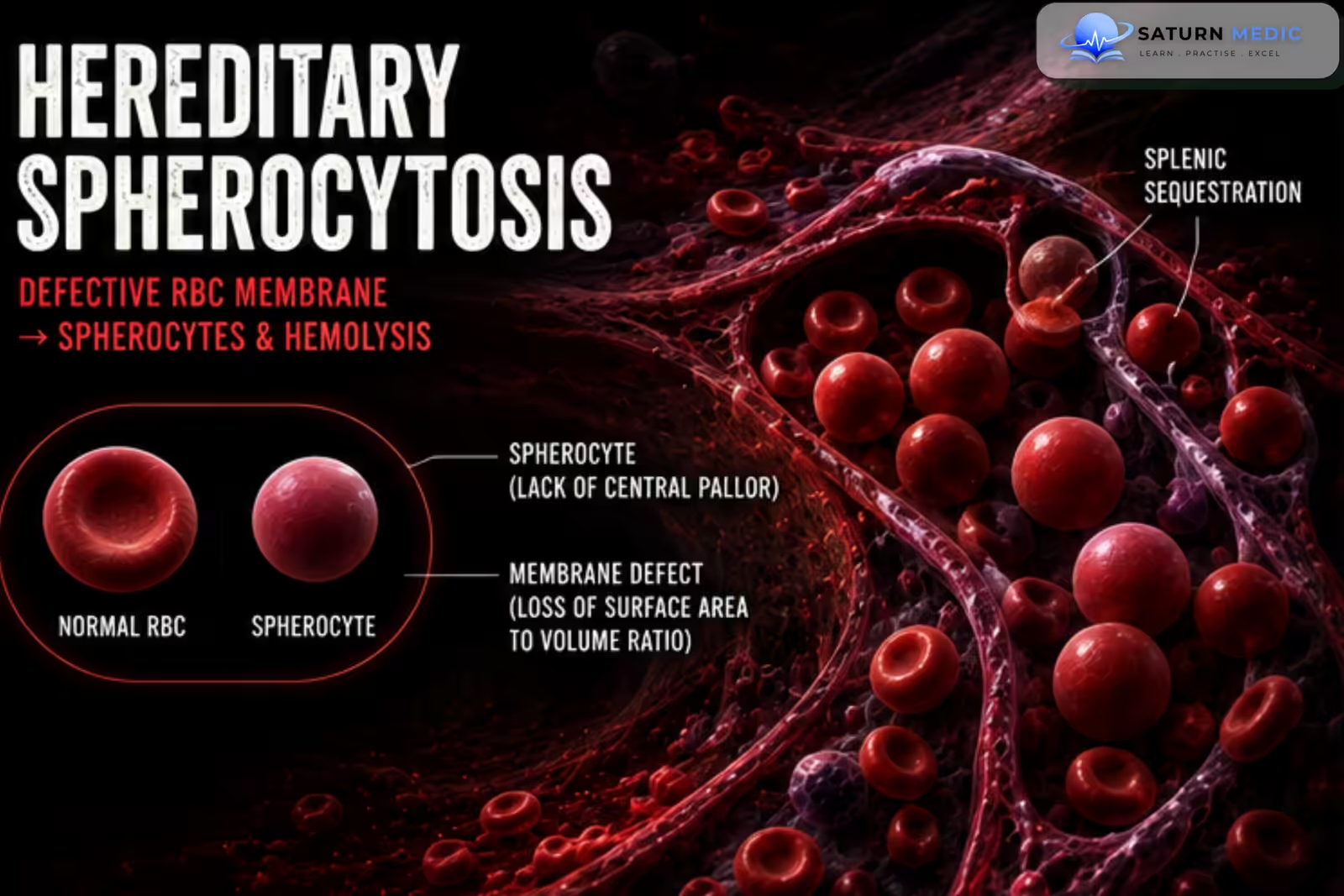
Hereditary Spherocytosis (HS) is the most common inherited red blood cell membrane disorder, characterized by the production of spherocytes that are fragile and prone to premature destruction in the spleen, resulting in chronic extravascular hemolytic anemia
Also called
ICD-10
Specialty
Onset
Reviewed
-
OverviewOverview
-
Etiology & Risk FactorsEtiology & Risk Factors
-
PathophysiologyPathophysiology
-
Clinical PresentationClinical Presentation
-
History TakingHistory Taking
-
Physical ExaminationPhysical Examination
-
InvestigationsInvestigations
-
DiagnosisDiagnosis
-
ManagementManagement
-
ComplicationsComplications
-
PrognosisPrognosis
-
Key Points / Clinical PearlsKey Points / Clinical Pearls
Overview
Hereditary Spherocytosis (HS) is caused by inherited defects in red blood cell membrane proteins such as spectrin, ankyrin, band 3, and protein 4.2. These defects produce spherical, poorly deformable red blood cells that become trapped and destroyed in the spleen, causing chronic hemolysis, anemia, jaundice, splenomegaly, and pigment gallstones.
Disease severity ranges from asymptomatic to severe transfusion-dependent anemia.
Etiology & Risk Factors
-Etiology
Hereditary Spherocytosis (HS) results from inherited mutations affecting red blood cell membrane proteins, including:
-
Ankyrin (ANK1)
-
Spectrin (α or β spectrin)
-
Band 3 protein
-
Protein 4.2
Inheritance
-
Autosomal dominant (≈75% of cases)
-
Autosomal recessive (less common)
-
Rare de novo mutations
-Risk Factors
-
Positive family history
-
Northern European ancestry
-
First-degree relative with HS
Pathophysiology
Inherited mutation in red blood cell membrane proteins → loss of membrane surface area → formation of rigid spherical red blood cells (spherocytes) → decreased deformability → trapping of spherocytes within splenic cords → splenic macrophage destruction (extravascular hemolysis) → chronic hemolytic anemia → increased bilirubin production → pigment gallstones → compensatory bone marrow erythroid hyperplasia with reticulocytosis
Clinical Presentation
-Symptoms
-
Fatigue
-
Weakness
-
Jaundice
-
Dark urine
-
Pallor
-
Exercise intolerance
-
Abdominal discomfort
-Signs
-
Splenomegaly
-
Pallor
-
Mild jaundice
-
Pigment gallstones
-
Growth delay (severe childhood disease)
-Acute Complications
-
Hemolytic crisis
-
Aplastic crisis (commonly triggered by Parvovirus B19)
-
Megaloblastic crisis (folate deficiency)
History Taking
-Ask about:
- Fatigue
- Jaundice
- Gallstone symptoms
- Dark urine
- Previous blood transfusions
- Family history of hemolytic anemia
- Splenectomy
- Episodes of aplastic crisis
- Recent viral illness
- Abdominal pain
- Growth and developmental history in children
Physical Examination
-General Examination
Look for:
-
Pallor
-
Jaundice
-
Tachycardia
-Abdominal Examination
Assess for:
-
Splenomegaly
-
Hepatomegaly (occasionally)
-
Right upper quadrant tenderness if gallstones are present
-Additional Examination
-
-
Growth assessment in children
-
Signs of chronic anemia
-
Investigations
-Complete Blood Count (CBC)
Typical findings:
-
Low hemoglobin (variable)
-
Increased MCHC (characteristic)
-
Normal or low MCV
-
Increased reticulocyte count
-Peripheral Blood Film
Characteristic findings:
-
Numerous spherocytes
-
Polychromasia
-
Reticulocytosis
-Hemolysis Screen
-
Increased indirect bilirubin
-
Increased LDH
-
Low haptoglobin
-
Increased reticulocyte count
-Direct Antiglobulin (Coombs) Test
-
Negative
Useful for distinguishing HS from autoimmune hemolytic anemia.
-Eosin-5-Maleimide (EMA) Binding Test
-
Preferred screening test
-
Reduced fluorescence supports the diagnosis.
-Osmotic Fragility Test
-
Increased osmotic fragility
-
Less commonly used now due to EMA testing
-Additional Investigations
-
-
Blood group and antibody screen (if transfusion required)
-
Serum folate
-
Abdominal ultrasound (splenomegaly or gallstones)
-
Genetic testing in selected cases
-
Diagnosis
Diagnosis is based on:
-
- Clinical features
- Family history
- Peripheral blood film showing spherocytes
- Evidence of hemolysis
- Negative Coombs test
- Positive EMA binding test or increased osmotic fragility
Management
-Mild Disease
-
Observation
-
Folic acid supplementation
-
Regular follow-up
-Moderate to Severe Disease
-
Blood transfusions when necessary
-
Folic acid supplementation
-
Monitor for hemolysis and gallstones
-Splenectomy
Consider in:
-
Moderate to severe symptomatic disease
-
Frequent transfusion requirements
-
Significant hemolysis
-
Poor quality of life
Usually delayed until after 5–6 years of age to reduce the risk of overwhelming post-splenectomy infection (OPSI).
-Cholecystectomy
Indicated for:
-
Symptomatic pigment gallstones
May be performed with splenectomy if appropriate.
Vaccination
Before splenectomy:
-
Pneumococcal vaccine
-
Meningococcal vaccine
-
Haemophilus influenzae type b (Hib) vaccine
Complications
- Chronic hemolytic anemia
- Pigment gallstones
- Aplastic crisis (Parvovirus B19)
- Hemolytic crisis
- Folate deficiency
- Splenic sequestration (rare)
- Iron overload (after repeated transfusions)
- Overwhelming post-splenectomy infection (OPSI)
Prognosis
The prognosis is excellent for most patients with appropriate management. Mild disease often requires no specific treatment, while splenectomy significantly improves anemia and reduces hemolysis in moderate to severe cases. Lifelong follow-up is recommended to monitor for gallstones and post-splenectomy complications.
Key Points / Clinical Pearls
- Hereditary Spherocytosis (HS) is the most common inherited red blood cell membrane disorder.
- It is usually inherited as an autosomal dominant condition.
- Defects in spectrin, ankyrin, band 3, or protein 4.2 produce spherocytes.
- Hemolysis is primarily extravascular and occurs in the spleen.
- The classic triad is anemia, jaundice, and splenomegaly.
- CBC often shows increased MCHC.
- Peripheral blood film demonstrates spherocytes.
- The EMA binding test is the preferred diagnostic test.
- The Coombs test is negative, helping distinguish HS from autoimmune hemolytic anemia.
- Splenectomy is the definitive treatment for selected patients with moderate to severe disease.
- National Center for Biotechnology Information (NIH). Hereditary Spherocytosis, StatPearls.
- Bolton-Maggs PH, Langer JC, Iolascon A, Tittensor P, King MJ; General Haematology Task Force of the British Committee for Standards in Haematology. Guidelines for the Diagnosis and Management of Hereditary Spherocytosis. Br J Haematol. 2012;156:37-49. Guideline Summary.
- Hereditary Spherocytosis: Linking Ion Transport Defects to Osmotic Gradient Ektacytometry Profiles - A Review. PMC12841313.
- An Overview of Hereditary Spherocytosis and the Curative Effects of Splenectomy. Front Physiol. Frontiers in Physiology.
- MedlinePlus, National Library of Medicine (NIH). Hereditary Spherocytosis: Medical Encyclopedia.