

Clinical Subject Page
Myelofibrosis (MF)
Myelofibrosis (MF) is a chronic myeloproliferative neoplasm (MPN) characterized by abnormal megakaryocyte proliferation, progressive bone marrow fibrosis, ineffective hematopoiesis, and extramedullary hematopoiesis. It commonly causes anemia, constitutional symptoms, and splenomegaly
Also called
ICD-10
Specialty
Onset
Reviewed
-
OverviewOverview
-
Etiology & Risk FactorsEtiology & Risk Factors
-
PathophysiologyPathophysiology
-
Clinical PresentationClinical Presentation
-
History TakingHistory Taking
-
Physical ExaminationPhysical Examination
-
InvestigationsInvestigations
-
DiagnosisDiagnosis
-
ManagementManagement
-
ComplicationsComplications
-
PrognosisPrognosis
-
Key Points / Clinical PearlsKey Points / Clinical Pearls
Overview
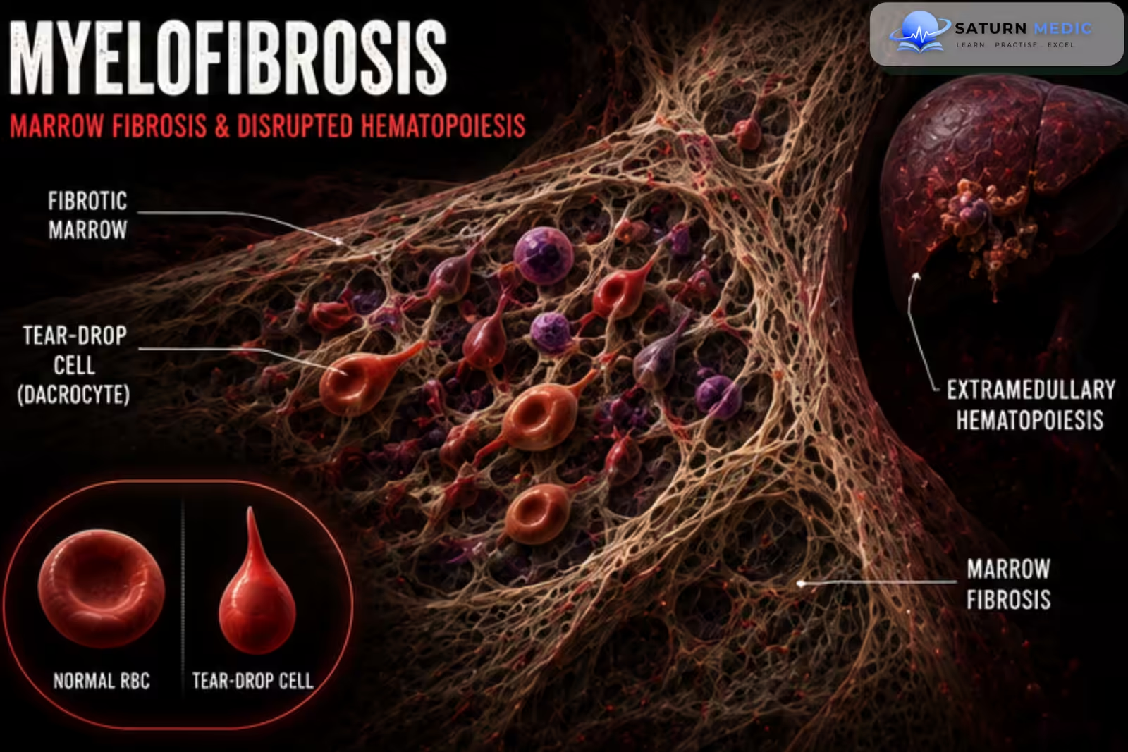
Myelofibrosis (MF) is a clonal myeloid disorder in which abnormal megakaryocytes release fibrogenic cytokines, causing progressive scarring of the bone marrow. Normal blood-cell production becomes impaired, resulting in anemia and other cytopenias. The body compensates by producing blood cells outside the bone marrow, especially in the spleen and liver, causing marked splenomegaly.
Etiology & Risk Factors
Myelofibrosis (MF) results from an acquired clonal abnormality of hematopoietic stem cells, commonly involving:
- JAK2
- CALR
- MPL
-Risk Factors
- Increasing age
- Male sex
- Previous myeloproliferative neoplasm
- Previous Polycythemia Vera (PV)
- Previous Essential Thrombocythemia (ET)
- Rare familial predisposition
Pathophysiology
Clonal mutation in hematopoietic stem cells → abnormal megakaryocyte proliferation → release of fibrogenic cytokines such as TGF-β and PDGF → progressive bone marrow fibrosis → ineffective hematopoiesis → anemia and other cytopenias → extramedullary hematopoiesis in the spleen and liver → massive splenomegaly and hepatomegaly → progressive bone marrow failure and possible transformation to AML.
Clinical Presentation
-Symptoms
Fatigue
Weakness
Weight loss
Fever
Night sweats
Early satiety
Abdominal fullness
Left upper quadrant discomfort
Bone pain
Dyspnea
-Signs
Massive splenomegaly
Hepatomegaly
Pallor
Cachexia
Petechiae or bruising
Signs of anemia
History Taking
-Ask about:
- Fatigue
- Weight loss
- Fever
- Night sweats
- Abdominal fullness
- Early satiety
- Bone pain
- Bleeding
- Previous thrombosis
- Previous PV or ET
Physical Examination
-General Examination
Look for:
Pallor
Weight loss
Cachexia
Petechiae
Ecchymoses
-Abdominal Examination
Assess for:
Massive splenomegaly
Hepatomegaly
-Additional Examination
Look for:
Signs of anemia
Bleeding manifestations
Lymphadenopathy (uncommon)
Investigations
Complete Blood Count (CBC)
Typical findings:
Anemia
Variable leukocyte count
Variable platelet count
Thrombocytopenia in advanced disease
Pancytopenia in severe disease
Peripheral Blood Film
Characteristic findings:
Teardrop cells (dacrocytes)
Leukoerythroblastic picture
Nucleated RBCs
Immature granulocytes
Abnormal platelets
Bone Marrow Aspiration & Biopsy
Typical findings:
Dry tap on aspiration
Hypercellularity in early disease
Megakaryocytic proliferation and atypia
Increased reticulin/collagen fibrosis
Molecular Testing
JAK2 mutation
CALR mutation
MPL mutation
Additional Investigations
LDH
Uric acid
Renal function
Liver function
Abdominal ultrasound or CT for splenomegaly
Cytogenetic testing
-Prognostic Assessment
Risk assessment may use:
DIPSS
DIPSS-Plus
MIPSS70
Diagnosis
-Diagnosis is based on:
- Bone marrow biopsy showing megakaryocytic proliferation and fibrosis
- JAK2, CALR, or MPL mutation when present
- Characteristic peripheral blood findings
- Splenomegaly and constitutional symptoms
- Exclusion of other myeloid neoplasms
Management
-Asymptomatic / Low-Risk Disease
Observation
Regular CBC and clinical monitoring
-Symptomatic Disease
Ruxolitinib
Fedratinib in selected patients
Treatment of anemia
Blood transfusions when required
-Anemia Management
Erythropoiesis-stimulating agents in selected patients
Transfusion support
Other anemia-directed therapy depending on the cause
-Severe Splenomegaly
JAK inhibitor therapy
Splenectomy in selected patients
Splenic irradiation rarely
-Curative Treatment
Allogeneic hematopoietic stem cell transplantation (HSCT)
Complications
- Severe anemia
- Massive splenomegaly
- Hepatomegaly
- Portal hypertension
- Extramedullary hematopoiesis
- Recurrent infections
- Bleeding
- Thrombosis
- Bone marrow failure
- Acute Myeloid Leukemia (AML) transformation
Prognosis
Prognosis varies according to disease risk, age, blood counts, symptoms, cytogenetic abnormalities, and molecular mutations. Some patients have an indolent course for many years, while high-risk disease may progress rapidly. Transformation to AML is associated with a poor prognosis.
Key Points / Clinical Pearls
- Myelofibrosis (MF) is a myeloproliferative neoplasm.
- Common mutations: JAK2, CALR, and MPL.
- Abnormal megakaryocytes cause progressive bone marrow fibrosis.
- Massive splenomegaly is characteristic.
- Peripheral blood shows teardrop cells and a leukoerythroblastic picture.
- Bone marrow aspiration may produce a dry tap.
- Bone marrow biopsy confirms fibrosis.
- Ruxolitinib is commonly used for symptomatic disease.
- Allogeneic HSCT is the only potentially curative treatment.
- MF can progress to bone marrow failure or AML.
- Thapa B, Fazal S, Parsi M, Rogers HJ. National Center for Biotechnology Information (NIH). Myeloproliferative Neoplasms, StatPearls.
- Kuykendall AT, Shah S, Talati C, et al. Myelofibrosis in 2019: Moving Beyond JAK2 Inhibition. Blood Cancer J. 2019;9:74. Blood Cancer Journal.
- JAK2, CALR, and MPL Mutation Profiles in BCR-ABL Negative Myeloproliferative Neoplasms, a Referral Center Experience in the Middle East. PMC8085288.
- Grinfeld J, Nangalia J, Baxter EJ, et al. Classification and Personalized Prognosis in Myeloproliferative Neoplasms. N Engl J Med. 2018;379:1416-1430.
- MedlinePlus, National Library of Medicine (NIH). Myelofibrosis: Medical Encyclopedia.