

Clinical Subject Page
Thalassemia
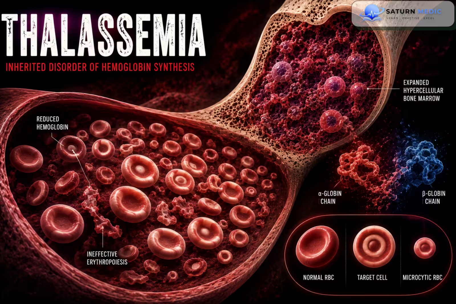
Thalassemia is a group of inherited hemoglobin disorders caused by reduced or absent production of alpha or beta globin chains. This leads to microcytic anemia, ineffective erythropoiesis, and increased red blood cell destruction
Also called
ICD-10
Specialty
Onset
Reviewed
-
OverviewOverview
-
Etiology & Risk FactorsEtiology & Risk Factors
-
PathophysiologyPathophysiology
-
Clinical PresentationClinical Presentation
-
History TakingHistory Taking
-
Physical ExaminationPhysical Examination
-
InvestigationsInvestigations
-
DiagnosisDiagnosis
-
ManagementManagement
-
ComplicationsComplications
-
PrognosisPrognosis
-
Key Points / Clinical PearlsKey Points / Clinical Pearls
Overview
Thalassemia occurs when the body produces insufficient amounts of one of the globin chains needed to make hemoglobin. The resulting red blood cells are small, pale, and easily destroyed, causing chronic anemia. Severe disease can lead to bone marrow expansion, hepatosplenomegaly, growth problems, and iron overload.
Etiology & Risk Factors
-Etiology
It is caused by inherited mutations or deletions in globin genes, resulting in reduced or absent production of globin chains.
–Alpha Thalassemia
Caused by abnormalities involving the alpha-globin genes on chromosome 16.
Severity depends mainly on the number of affected alpha-globin genes.
–Beta Thalassemia
Caused by mutations affecting the beta-globin gene on chromosome 11.
Mutations may cause:
Reduced beta-globin production
Absent beta-globin production
-Types
Alpha Thalassemia
Silent carrier
Alpha thalassemia trait
Hemoglobin H disease
Hydrops fetalis with Hb Bart’s
Beta Thalassemia
Beta thalassemia trait
Beta thalassemia intermedia
Beta thalassemia major
-Risk Factors
Family history
Parents carrying thalassemia mutations
Consanguinity
Mediterranean ancestry
Middle Eastern ancestry
South Asian ancestry
Southeast Asian ancestry
African ancestry
Pathophysiology
Globin gene mutation → reduced globin-chain synthesis → imbalance between globin chains → ineffective hemoglobin production → ineffective erythropoiesis and red blood cell destruction → chronic anemia → increased erythropoietin production → bone marrow expansion and extramedullary hematopoiesis → hepatosplenomegaly and skeletal changes → chronic anemia and transfusion therapy may lead to iron overload.
Clinical Presentation
-Symptoms
Mild disease may be asymptomatic.
Moderate or severe disease may cause:
Fatigue
Weakness
Pallor
Shortness of breath
Dizziness
Poor growth
Delayed puberty
Abdominal fullness
Exercise intolerance
-Signs
Pallor
Jaundice
Hepatomegaly
Splenomegaly
Growth retardation
Skeletal abnormalities in severe untreated disease
Signs of iron overload in chronically transfused patients
-Severe Disease
Beta thalassemia major may present during infancy with:
Severe anemia
Failure to thrive
Feeding difficulties
Recurrent infections
Marked hepatosplenomegaly
History Taking
-Ask about:
- Fatigue
- Pallor
- Exercise intolerance
- Jaundice
- Recurrent infections
- Growth and development
- Delayed puberty
- Abdominal fullness
- Previous blood transfusions
- Iron chelation therapy
- Family history
- Consanguinity
- Previous diagnosis of anemia
- Dietary history
- Previous iron therapy
Physical Examination
-General Examination
Look for:
Pallor
Jaundice
Growth retardation
Delayed puberty
Signs of chronic anemia
-Abdominal Examination
Assess for:
Splenomegaly
Hepatomegaly
-Skeletal Examination
In severe untreated disease, look for:
Frontal bossing
Maxillary enlargement
Facial bone changes
Bone deformities
-Cardiovascular Examination
Assess for:
Tachycardia
Flow murmur
Signs of heart failure
Cardiac complications from iron overload
Investigations
-Complete Blood Count
Typical findings:
Low hemoglobin
Marked microcytosis
Low MCV
Low MCH
RBC count may be relatively high, particularly in thalassemia trait
Variable reticulocytosis
-Peripheral Blood Film
May show:
Microcytosis
Hypochromia
Target cells
Anisopoikilocytosis
Basophilic stippling
Nucleated red blood cells in severe disease
-Iron Studies
Usually:
Ferritin normal or increased
Serum iron normal or increased
–Iron studies are important to distinguish thalassemia from iron deficiency anemia.
-Hemoglobin Analysis
Hemoglobin Electrophoresis / HPLC: Useful for identifying abnormal hemoglobin patterns.
In beta thalassemia trait:
HbA2 is usually increased
HbF may be mildly increased
In severe beta thalassemia:
HbF is markedly increased
HbA may be reduced or absent depending on the genotype
Alpha thalassemia may have a normal adult hemoglobin electrophoresis, particularly in the trait state.
-Genetic Testing
Useful for:
Confirming the diagnosis
Identifying specific mutations
Prenatal diagnosis
Family screening
Additional Investigations in Severe Disease
Liver function tests
Ferritin
Transferrin saturation
Cardiac MRI for iron overload
Liver MRI for iron concentration
Endocrine assessment
Bone density assessment
Diagnosis
-Diagnosis is based on:
- Chronic microcytic anemia
- Peripheral blood film findings
- Iron studies
- Hemoglobin electrophoresis or HPLC
- Genetic testing when required
- Family history
Management
-Thalassemia Trait
Usually requires:
No specific treatment
Avoid unnecessary iron therapy
Genetic counseling
Screening of partners when appropriate
-Severe Thalassemia
1. Regular Blood Transfusion
Used for severe anemia and transfusion-dependent disease.
Goals include:
Maintaining adequate hemoglobin
Suppressing ineffective erythropoiesis
Reducing skeletal complications
Supporting normal growth and development
2. Iron Chelation
Repeated transfusions cause iron overload.
Chelation options include:
Deferasirox
Deferoxamine
Deferiprone
Treatment is guided by iron burden and organ involvement.
3. Folic Acid
May be used in patients with increased erythropoiesis when clinically appropriate.
4. Splenectomy
May be considered in selected patients with:
Severe hypersplenism
Increasing transfusion requirements
Symptomatic splenomegaly
5. Luspatercept
May be used in selected adults with transfusion-dependent beta thalassemia to reduce transfusion requirements.
6. Hematopoietic Stem Cell Transplantation
Allogeneic hematopoietic stem cell transplantation can be curative, particularly when performed in appropriately selected younger patients.
7. Gene Therapy
Gene-based therapies are available for selected patients with transfusion-dependent beta thalassemia in appropriate specialist settings.
Complications
- Severe chronic anemia
- Growth retardation
- Delayed puberty
- Hepatosplenomegaly
- Skeletal abnormalities
- Gallstones
- Extramedullary hematopoiesis
- Iron overload
- Cardiomyopathy
- Liver fibrosis
- Endocrine dysfunction
- Diabetes mellitus
- Hypogonadism
- Osteoporosis
- Increased infection risk
- Transfusion reactions
- Alloimmunization
Prognosis
Prognosis varies according to the type and severity of thalassemia. Patients with thalassemia trait usually have a normal life expectancy, while severe transfusion-dependent disease requires lifelong specialist care. Modern transfusion therapy, iron chelation, stem cell transplantation, and newer gene-based treatments have significantly improved outcomes.
Key Points / Clinical Pearls
- Thalassemia is an inherited hemoglobin disorder.
- It results from reduced production of alpha or beta globin chains.
- It causes microcytic, hypochromic anemia.
- Thalassemia trait is often mild or asymptomatic.
- Severe disease causes chronic anemia and hepatosplenomegaly.
- The red blood cell count may be relatively high despite microcytosis.
- Iron studies are usually normal unless iron deficiency is also present.
- Hemoglobin electrophoresis is useful, particularly for beta thalassemia.
- Genetic testing is important in selected cases.
- Severe disease may require regular blood transfusions.
- Repeated transfusions can cause iron overload.
- Iron chelation is essential when significant iron overload develops.
- Hematopoietic stem cell transplantation can be curative in suitable patients.
- National Center for Biotechnology Information (NIH). Thalassemia, StatPearls.
- Needs T, Gonzalez-Mosquera LF, Lynch DT. National Center for Biotechnology Information (NIH). Beta Thalassemia, StatPearls.
- Cappellini MD, Farmakis D, Porter J, Taher A, et al. Thalassemia International Federation (TIF) Guidelines for the Management of Transfusion-Dependent Beta-Thalassemia. 4th ed. PMC11880825.
- Harewood J, Azevedo AM. National Center for Biotechnology Information (NIH). Alpha Thalassemia, StatPearls.
- MedlinePlus, National Library of Medicine (NIH). Thalassemia: Health Topic.