

Clinical Subject Page
Pleural Effusion
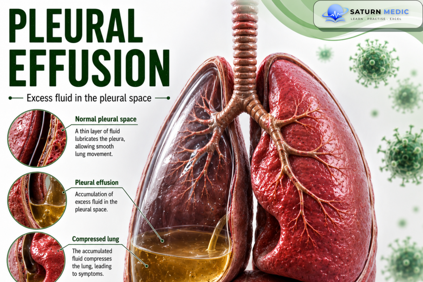
It is the abnormal accumulation of fluid within the pleural cavity (between the visceral and parietal pleura
Also called
ICD-10
Specialty
Onset
Reviewed
-
OverviewOverview
-
Etiology & Risk FactorsEtiology & Risk Factors
-
PathophysiologyPathophysiology
-
Clinical PresentationClinical Presentation
-
History TakingHistory Taking
-
Physical ExaminationPhysical Examination
-
InvestigationsInvestigations
-
DiagnosisDiagnosis
-
ManagementManagement
-
ComplicationsComplications
-
PrognosisPrognosis
-
Key Points / Clinical PearlsKey Points / Clinical Pearls
Overview
Normally, a small amount of pleural fluid lubricates the pleural surfaces and facilitates
lung movement. Excess fluid accumulates when hydrostatic, oncotic, permeability, or
lymphatic drainage mechanisms are disrupted. Pleural effusions are classified as
transudative or exudative based on their underlying mechanism and pleural fluid
analysis.
Etiology & Risk Factors
-Transudative pleural effusion
Caused by:
• Increased capillary hydrostatic pressure
• Decreased capillary oncotic pressure
Common causes:
• Congestive heart failure
• Low-protein states (e.g., chronic kidney disease, liver disease)
-Exudative pleural effusion
Caused by:
• Increased capillary permeability (e.g., inflammation)
Common causes:
• Pulmonary infections
• Pulmonary or metastatic malignancy
• Pancreatitis
• Autoimmune diseases
• Hemothorax
• Chylothorax
• Pulmonary embolism
Pathophysiology
It develops when the balance between pleural fluid production and
removal is disrupted.
Transudative effusion
• Increased hydrostatic pressure
• Decreased oncotic pressure
Exudative effusion
• Increased vascular permeability due to inflammation or injury
• Leakage of protein-rich fluid into the pleural space
Clinical Presentation
- Clinical Presentation:
Presentation varies depending on:
• Size of the effusion
• Underlying disease
• Presence of respiratory compromise
Red flag features include:
• Respiratory distress
• Hemodynamic instability
• Complete lung opacification
• Mediastinal shift
• Tracheal deviation
• Sepsis
History Taking
Assess for:
• Recent respiratory infection
• Fever
• Weight loss
• Known cardiac, renal, or liver disease
• Malignancy
• Recent changes in medical conditions or treatment
Physical Examination
Assess:
• Respiratory status
• Hemodynamic stability
• Oxygen saturation
• Signs of respiratory distress
Perform an ABCDE assessment in unstable patients
Investigations
-Imaging :
• Chest X-ray
• Thoracic ultrasound
• Chest CT (especially for very small effusions)
Pleural fluid evaluation
• Diagnostic thoracentesis
• Pleural fluid analysis
Light’s Criteria
To Determine if the effusion is exudative if any of the following are present:
• Pleural fluid protein/serum protein ratio >0.5
• Pleural fluid LDH/serum LDH ratio >0.6
• Pleural fluid LDH >⅔ the upper limit of normal serum LDH
Transudative effusions have values below these thresholds.
Diagnosis
-Diagnosis is based on:
• Clinical assessment
• Chest imaging
• Diagnostic thoracentesis with pleural fluid analysis
• Classification into transudative or exudative effusion using Light’s criteria
Related Topics
Management of Pleural Effusion
-Unstable patients :
•Begin respiratory support
• Perform urgent therapeutic thoracentesis
• Oxygen therapy as needed
• Continuous cardiac monitoring
• Continuous pulse oximetry
• ICU admission if hemodynamically unstable
-Stable patients:
• Identify and treat the underlying condition
• Consider elective therapeutic thoracentesis for symptom relief
• Consider specialized procedures when indicated:
• Tube thoracostomy
• Indwelling pleural catheter
• Pleurodesis
• Surgical management after specialty consultation
Disposition depends on:
• Stability
• Underlying diagnosis
• Effusion size
• Response to treatment
Complications of Pleural Effusion
• Respiratory distress
• Respiratory failure
• Hemodynamic instability
• Empyema
• Massive hemothorax
• Malignant pleural effusion with respiratory failure
Prognosis of Pleural Effusion
- Prognosis depends largely on the underlying cause and the patient’s response to treatment
- Management focuses primarily on identifying and treating the underlying
disease.
Key Points / Clinical Pearls of Pleural Effusion
- • it is the abnormal fluid accumulation in the pleural cavity.
• Classified as transudative or exudative.
• Chest X-ray and ultrasound are primary imaging modalities.
• CT may detect very small effusions.
• Thoracentesis is both diagnostic and therapeutic.
• Light’s criteria differentiate transudates from exudates.
• Treat the underlying cause.
- Krishna R, Antoine MH, Alahmadi MH, Rudrappa M. National Center for Biotechnology Information (NIH). Pleural Effusion, StatPearls.
- MedlinePlus, National Library of Medicine (NIH). Pleural Effusion: Medical Encyclopedia.
- Light RW, Macgregor MI, Luchsinger PC, Ball WC Jr. Pleural Effusions: The Diagnostic Separation of Transudates and Exudates. Ann Intern Med. 1972. PMID: 4642731.
- D'Agostino HP, Edens MA. National Center for Biotechnology Information (NIH). Physiology, Pleural Fluid, StatPearls.
- Sharma S, Arora RD, Boster J. National Center for Biotechnology Information (NIH). Malignant Pleural Effusion, StatPearls.