

Clinical Subject Page
Spontaneous Pneumothorax
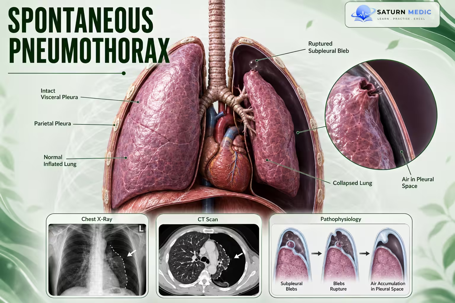
Spontaneous pneumothorax is the presence of air within the pleural space causing
partial or complete collapse of the lung without preceding trauma
Also called
ICD-10
Specialty
Onset
Reviewed
-
OverviewOverview
-
Etiology & Risk FactorsEtiology & Risk Factors
-
PathophysiologyPathophysiology
-
Clinical PresentationClinical Presentation
-
History TakingHistory Taking
-
Physical ExaminationPhysical Examination
-
InvestigationsInvestigations
-
DiagnosisDiagnosis
-
ManagementManagement
-
ComplicationsComplications
-
PrognosisPrognosis
-
Key Points / Clinical PearlsKey Points / Clinical Pearls
Overview
It is classified into:
• Primary spontaneous pneumothorax (PSP): Occurs in patients without clinically
apparent underlying lung disease.
• Secondary spontaneous pneumothorax (SSP): Occurs as a complication of underlying
lung disease.
• Recurrent pneumothorax refers to a second spontaneous episode (ipsilateral or
contralateral).
Etiology & Risk Factors
-Primary spontaneous pneumothorax :
• No clinically apparent underlying lung disease.
-Secondary spontaneous pneumothorax :
• Occurs due to underlying lung disease.
-Higher-risk patients :
• Age >50 years
• Smoking history
• Recurrent disease
Pathophysiology
• Air accumulates in the pleural space.
• Loss of negative intrapleural pressure leads to partial or complete lung collapse.
• Progressive accumulation of air may produce tension pneumothorax with
cardiorespiratory compromise.
Clinical Presentation
- Clinical features vary depending on:
• Size
• Patient stability
• Presence of underlying lung disease
• Presence of tension physiology
Patients are considered stable only if ALL of the following are present:
• Respiratory rate <24/min
• SpO₂ >90% on room air
• Able to speak in complete sentences
• Heart rate 60–120/min
• Normal blood pressure
Failure to meet any criterion indicates an unstable patient.
History Taking
-
Assess for:
• Sudden onset symptoms
• Previous spontaneous pneumothorax
• Smoking history
• Underlying lung disease
• Recurrence
• Symptoms suggesting respiratory compromise
Physical Examination
Assess:
• Respiratory rate
• Heart rate
• Blood pressure
• Oxygen saturation
• Ability to speak in full sentences
• Signs of respiratory distress
• Features suggesting tension pneumothorax
Investigations
- Chest X-ray
• Repeat chest X-ray after:
• 3–6 hours for small stable primary spontaneous pneumothorax
• Chest tube insertion
-Assess The size using apex-to-cupola distance :
• <3 cm Small
• ≥3 cm Large
Diagnosis
- Diagnosis is based on:
• Clinical assessment
• Chest imaging
Tension pneumothorax remains a clinical diagnosis requiring immediate treatment.
Related Topics
Management of Spontaneous Pneumothorax
-Initial management for all patients :
• Continuous telemetry
• Continuous pulse oximetry
• Supplemental oxygen (if no risk factors for hypercapnia; target SpO₂ ≥96%)
• Avoid positive-pressure ventilation
• If mechanical ventilation is required, perform tube thoracostomy first
-Unstable patients :
• Immediate needle thoracostomy for suspected tension pneumothorax
• Follow immediately with chest tube placement
• Consider finger thoracostomy if needle decompression fails
• ICU transfer
• Thoracic surgery consultation
-Stable Primary Spontaneous Pneumothorax :
Small (<3 cm)
• Observation
• Repeat chest X-ray after 3–6 hours
• If stable/improving → consider outpatient management
• If enlarging → chest tube
Large (≥3 cm)
• Chest tube placement (small-bore catheter or chest tube)
• Needle aspiration may be considered
• Conservative management may be considered in carefully selected healthy patients
with:
• Initial episode
• No respiratory distress
• No progression on repeat imaging
-Stable Secondary Spontaneous Pneumothorax :
Small (<3 cm)
• Admission for monitoring
• Observation or chest tube placement
• Thoracic surgery consultation
Large (≥3 cm)
• Chest tube placement
• ICU consideration
• Thoracic surgery consultation
Complications of Spontaneous Pneumothorax
• Recurrece
• Tension pneumothorax
• Cardiorespiratory compromise
Prognosis of Spontaneous Pneumothorax
- Depends on:
• Primary vs secondary spontaneous pneumothorax
• Pneumothorax size
• Patient stability
• Prompt recognition and management
Small stable primary spontaneous pneumothoraces often resolve spontaneously within
approximately 10 days
Key Points / Clinical Pearls of Spontaneous Pneumothorax
- • Air in pleural space causes lung collapse.
• Primary = no clinically apparent lung disease.
• Secondary = underlying lung disease.
• Assess stability first.
• Chest X-ray confirms diagnosis in stable patients.
• Tension pneumothorax is a clinical diagnosis.
- Gurung I, Ghassemzadeh S. National Center for Biotechnology Information (NIH). Spontaneous Pneumothorax, StatPearls.
- MedlinePlus, National Library of Medicine (NIH). Pneumothorax: Medical Encyclopedia.
- Roberts ME, Rahman NM, Maskell NA, et al. British Thoracic Society Guideline for Pleural Disease. Thorax. 2023. PMID: 37553157.
- Sankari A, Gupta N. National Center for Biotechnology Information (NIH). Acute Pneumothorax Evaluation and Treatment, StatPearls.
- Mummadi SR, de Longpre J, Hahn PY. Comparative Effectiveness of Interventions in Initial Management of Spontaneous Pneumothorax: A Systematic Review and Bayesian Network Meta-Analysis. Ann Emerg Med. 2020. PMID: 32115228.