

Clinical Subject Page
Tension Pneumothorax
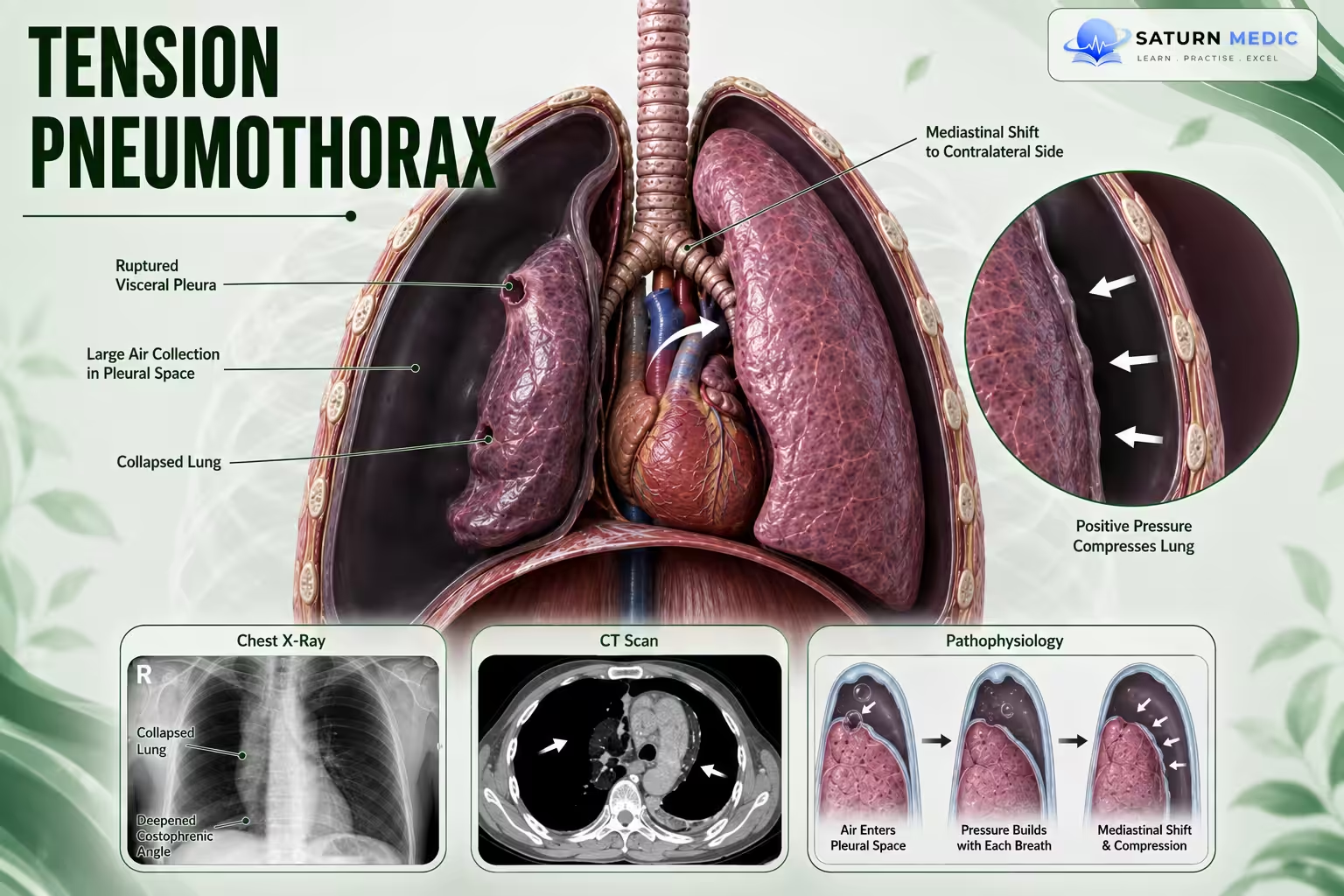
Tension Pneumothorax is A life-threatening form of pneumothorax in which progressively increasing pressure within the pleural space causes severe respiratory and cardiovascular compromise. It is a clinical diagnosis that requires immediate treatment.
ICD-10
Specialty
Onset
Reviewed
-
OverviewOverview
-
Etiology & Risk FactorsEtiology & Risk Factors
-
PathophysiologyPathophysiology
-
Clinical PresentationClinical Presentation
-
History TakingHistory Taking
-
Physical ExaminationPhysical Examination
-
InvestigationsInvestigations
-
DiagnosisDiagnosis
-
ManagementManagement
-
ComplicationsComplications
-
PrognosisPrognosis
-
Key Points / Clinical PearlsKey Points / Clinical Pearls
Overview
A one-way valve mechanism allows air to enter the pleural space during inspiration but
prevents it from escaping. Progressive pressure compresses the lungs and mediastinal
structures, leading to impaired ventilation, decreased venous return, reduced cardiac
output, hypoxia, and hemodynamic instability.
Etiology & Risk Factors
Causes
• Disruption of the:
• Visceral pleura
• Parietal pleura
• Tracheobronchial tree
May occur as:
• Complication of spontaneous pneumothorax
• Traumatic pneumothorax
Pathophysiology
Pathophysiology:
1. Disruption of the pleura or tracheobronchial tree.
2. One-way valve mechanism develops.
3. Air enters the pleural space during inspiration but cannot escape.
4. Progressive accumulation of intrapleural air increases intrathoracic pressure.
5. Collapse of the ipsilateral lung.
6. Compression of:
• Contralateral lung
• Trachea
• Heart
• Superior vena cava
• Angulation of the inferior vena cava
7. Reduced venous return.
8. Reduced cardiac output.
9. Hypoxia and hemodynamic instability.
Clinical Presentation
Patients May Develop
-
Respiratory compromise
-
Hypoxia
-
Hemodynamic instability
An Unstable Patient May Have
-
Respiratory rate >24/min
-
SpO₂ <90% on room air
-
Heart rate <60 or >120/min
-
Hypotension
-
Inability to speak in complete sentences due to dyspnea
History Taking
-Assess for:
• Sudden onset respiratory symptoms
• Recent spontaneous pneumothorax
• Chest trauma
• Rapid clinical deterioration
Physical Examination
Assess:
• Respiratory rate
• Heart rate
• Blood pressure
• Oxygen saturation
• Ability to speak in complete sentences
• Signs of respiratory distress
• Hemodynamic instability
Investigations
• Diagnosis is primarily clinical.
• Serial chest X-rays after emergency treatment.
• Continuous pulse oximetry.
• Continuous telemetry
Diagnosis
Diagnosis is based on:
• Clinical evidence of progressive respiratory and cardiovascular compromise.
• Imaging should not delay emergency treatment.
Related Topics
Management of Tension Pneumothorax
Immediate management
• Administer 100% FiO₂ (high-concentration supplemental oxygen).
• Avoid positive-pressure ventilation.
• Perform emergency needle decompression in hemodynamically unstable patients.
• Usually in the 5th intercostal space, anterior to the midaxillary line.
• Immediately follow with tube thoracostomy (chest tube).
• Continuous telemetry.
• Continuous pulse oximetry.
• Serial chest X-rays after decompression.
• ICU transfer
Complications of Tension Pneumothorax
• Ipsilateral lung collapse
• Compression of the contralateral lung
• Compression of the heart
• Compression of the superior vena cava
• Reduced venous return
• Reduced cardiac output
• Severe hypoxia
• Hemodynamic instability
Prognosis of Tension Pneumothorax
- Prognosis depends on rapid recognition and immediate decompression.
- Delayed
treatment may rapidly result in severe cardiorespiratory compromise.
Key Points / Clinical Pearls of Tension Pneumothorax
• Medical emergency.
• Caused by a one-way valve mechanism.
• Air enters but cannot leave the pleural space.
• Progressive pressure compresses thoracic organs.
• Causes hypoxia and hemodynamic instability.
• Diagnosis is clinical.
• Never delay treatment for imaging.
• Immediate needle decompression followed by chest tube insertion.
• Administer 100% oxygen.
• ICU monitoring after stabilization.
- Jalota Sahota R, Sayad E. National Center for Biotechnology Information (NIH). Tension Pneumothorax, StatPearls.
- MedlinePlus, National Library of Medicine (NIH). Pneumothorax: Medical Encyclopedia.
- Sankari A, Gupta N. National Center for Biotechnology Information (NIH). Acute Pneumothorax Evaluation and Treatment, StatPearls.
- Inaba K, Ives C, McClure K, et al. Optimal Positioning for Emergent Needle Thoracostomy: A Cadaver-Based Study. J Trauma. 2011. PMID: 22071923.
- Laan DV, Vu TD, Thiels CA, et al. Chest Wall Thickness and Decompression Failure: A Systematic Review and Meta-Analysis Comparing Anatomic Locations in Needle Thoracostomy. Injury. 2016. PMID: 26724173.