

Clinical Subject Page
Superior Vena Cava (SVC) Syndrome
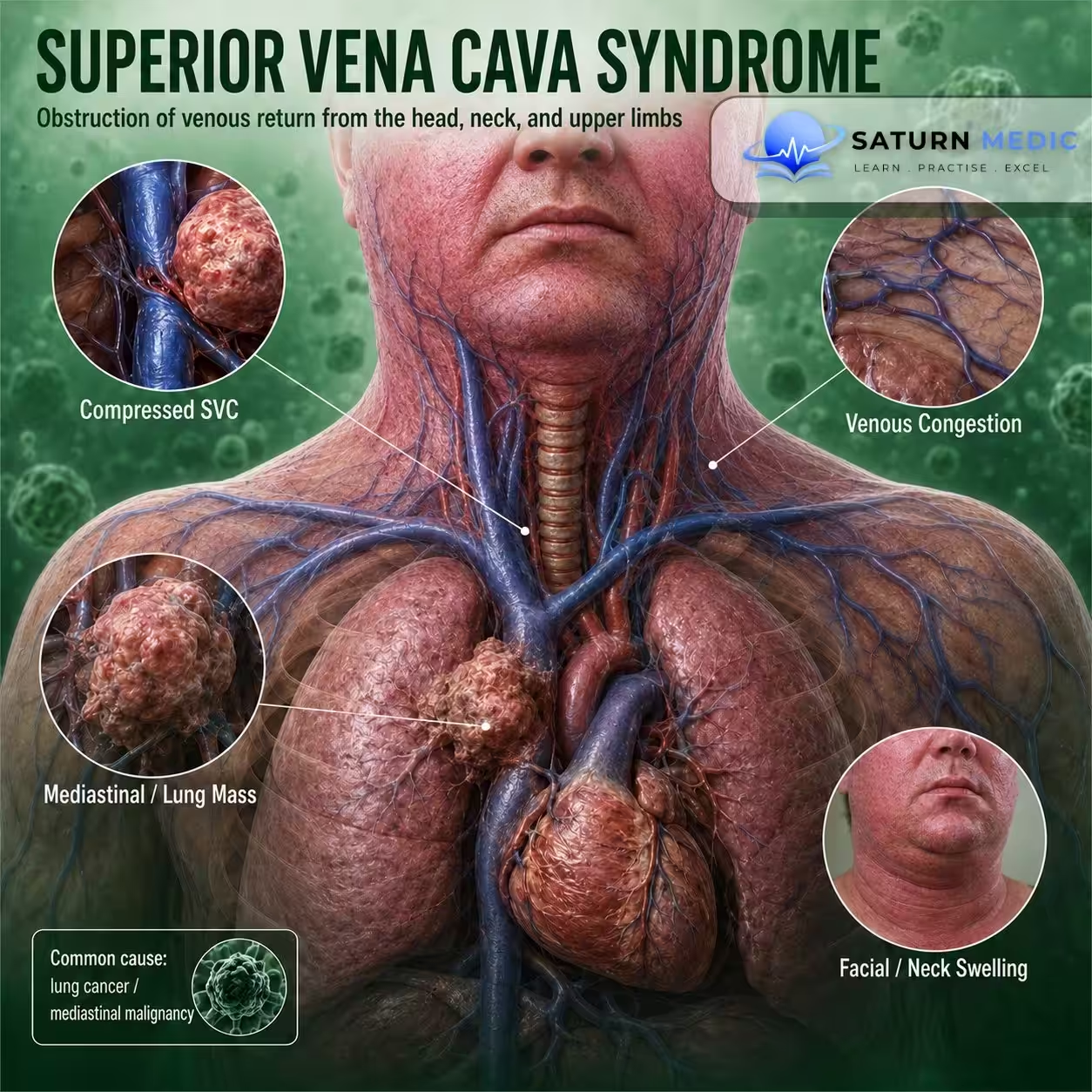
Superior vena cava (SVC) syndrome is venous congestion of the head, neck, and upper extremities caused by impaired blood flow through the superior vena cava to the right atrium.
Also called
ICD-10
Specialty
Onset
Reviewed
-
OverviewOverview
-
Etiology & Risk FactorsEtiology & Risk Factors
-
PathophysiologyPathophysiology
-
Clinical PresentationClinical Presentation
-
History TakingHistory Taking
-
Physical ExaminationPhysical Examination
-
InvestigationsInvestigations
-
DiagnosisDiagnosis
-
ManagementManagement
-
ComplicationsComplications
-
PrognosisPrognosis
-
Key Points / Clinical PearlsKey Points / Clinical Pearls
Overview
- Superior Vena Cava (SVC) Syndrome results from obstruction of the superior vena cava, either by external compression or intraluminal obstruction. Malignancy is the most common cause. Patients may present with facial swelling, upper extremity edema, dyspnea, and venous distension. Severe cases require emergency treatment.
Etiology & Risk Factors
Malignant causes (most common):
Responsible for >80% of cases:
• Non-small cell lung cancer (NSCLC)
• Small cell lung cancer (SCLC)
• Non-Hodgkin lymphoma (NHL)
Less common:
• Metastatic breast cancer
• Germ cell tumors
• Thymoma
• Mesothelioma
Nonmalignant causes
Most commonly:
• Catheter-associated thrombosis
• Pacemaker wire-associated thrombosis
Other causes:
• Fibrosis after radiotherapy or chemotherapy
• Tuberculosis
• Tertiary syphilis
• Sarcoidosis
• Goiter
• Aortic aneurysm
• Pancoast tumors and other mediastinal masses causing extrinsic compression
Pathophysiology
• Obstruction of the superior vena cava by:
• Intraluminal thrombosis
• Tumor invasion
• External compression
• Impaired venous return from the head, neck, and upper extremities
• Venous congestion
• Development of collateral veins if obstruction develops gradually
Clinical Presentation
Venous congestion
• Facial edema (facial plethora)
• Upper extremity edema
• Prominent veins over the chest, face, and upper limbs
• Jugular venous distension
Respiratory and neck symptoms
• Dyspnea
• Cough
• Hoarseness
• Stridor
• Dysphagia
Neurological symptoms
• Headache
• Dizziness
• Confusion
• Mental obtundation
• Visual impairment
Other
• Orthostatic hypotension
• Syncope
• Renal failure
History Taking
Assess for:
• Facial swelling
• Arm swelling
• Dyspnea
• Cough
• Hoarseness
• Dysphagia
• Headache
• Visual symptoms
• Syncope
• History of malignancy
• Indwelling central venous catheter or pacemaker
Physical Examination
Assess for:
• Facial plethora
• Upper limb edema
• Dilated chest wall veins
• Jugular venous distension
• Respiratory distress
• Hoarseness
• Stridor
• Neurological deficits
• Signs of raised intracranial pressure
Investigations
Unstable patients
• Invasive venography (gold standard)
• Endovascular assessment ± stent placement
Stable patients
Imaging
• CT chest with CT venography (modality of choice)
Findings:
• SVC obstruction
• Dilated collateral veins
• Underlying tumor or other cause
Doppler ultrasound
If catheter-associated thrombosis is suspected.
Additional imaging
• MRI chest with MR venography (if CT contraindicated)
• Chest X-ray (may show mediastinal enlargement, hilar lymphadenopathy, pleural effusion)
Diagnosis
Diagnosis is based on:
• Clinical features
• CT chest with CT venography
• Invasive venography in unstable patients
• Identification of the underlying cause
Related Topics
Management
General principles
• Treat the underlying cause.
• Early specialist involvement:
• Oncology
• Radiation oncology
• Vascular surgery
• Interventional radiology
Supportive treatment
• Elevate the head
• Oxygen therapy
• Pain management
• Consider fluid restriction
• Consider loop diuretics
• Remove intravascular catheter if catheter-associated thrombosis is present
Emergency treatment
For severe or life-threatening symptoms:
• Emergency endovascular treatment
• Venography with stent placement
Medical treatment
Malignant Superior Vena Cava (SVC) Syndrome
After histologic confirmation:
• Radiotherapy (e.g., radiosensitive tumors such as NSCLC)
• Chemotherapy (e.g., SCLC, NHL, germ cell tumors)
Glucocorticoids
Indications:
• Steroid-responsive tumors (e.g., NHL, thymoma)
• Laryngeal edema
• Prevention of radiation-induced edema
Example:
• Dexamethasone
Antithrombotic therapy
For thrombotic Superior Vena Cava (SVC) Syndrome:
• Initial parenteral anticoagulation (e.g., enoxaparin)
• Consider thrombolysis in selected patients
Surgical treatment
Selected patients:
• Surgical bypass
• SVC reconstruction
Complications
• Cerebral edema
• Laryngeal edema
• Increased intracranial pressure
• Hemodynamic instability
• Syncope
• Respiratory compromise
Prognosis
- Prognosis depends primarily on the underlying cause and the speed of diagnosis and treatment. Malignant SVC syndrome generally requires tumor-directed therapy, whereas thrombotic disease requires anticoagulation and/or endovascular intervention.
Key Points / Clinical Pearls
• Superior Vena Cava (SVC) Syndrome = obstruction of venous return from the head, neck, and upper extremities.
• Most commonly caused by malignancy.
• NSCLC, SCLC, and NHL account for >80% of cases.
• Facial edema and upper limb swelling are classic findings.
• CT chest with CT venography is the investigation of choice in stable patients.
• Venography with stent placement is indicated for severe or unstable cases.
• Treat the underlying cause.
• Severe Superior Vena Cava (SVC) Syndrome is an oncologic emergency.
- Seligson MT, Surowiec SM. National Center for Biotechnology Information (NIH). Superior Vena Cava Syndrome, StatPearls.
- White HJ, Soos MP. National Center for Biotechnology Information (NIH). Anatomy, Thorax, Superior Vena Cava, StatPearls.
- MedlinePlus, National Library of Medicine (NIH). Superior Vena Cava Obstruction: Medical Encyclopedia.
- Azizi AH, Shafi I, Shah N, et al. Superior Vena Cava Syndrome. JACC Cardiovasc Interv. 2020. PMID: 32973835.
- Rice TW, Rodriguez RM, Light RW. The Superior Vena Cava Syndrome: Clinical Characteristics and Evolving Etiology. Medicine (Baltimore). 2006. PMID: 16609349.