

Clinical Subject Page
Helminthic infection
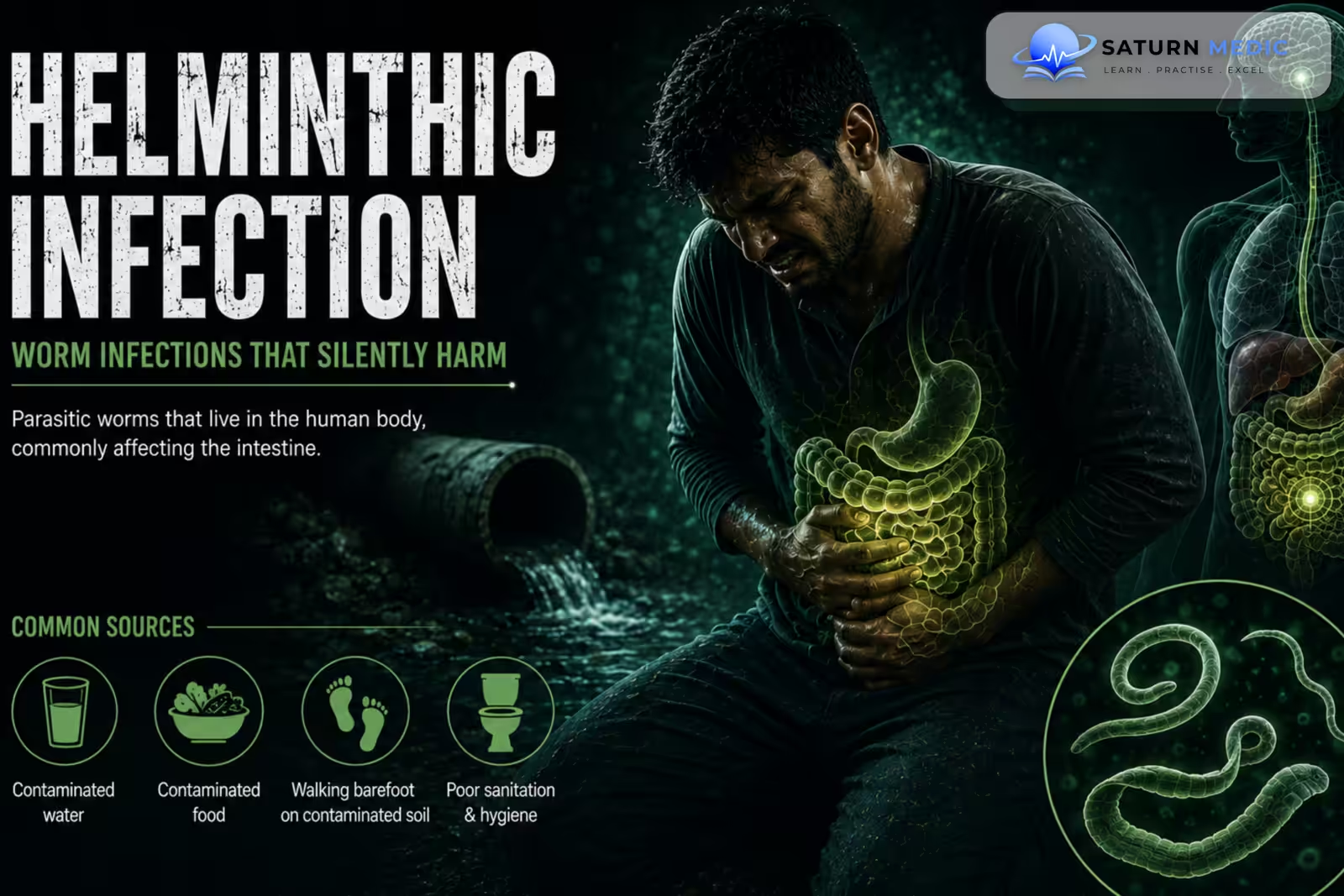
Helminthic Infection refers to disease caused by parasitic worms that infect humans. Helminths are broadly classified into nematodes (roundworms), cestodes (tapeworms), and trematodes (flukes). Clinical manifestations range from mild intestinal symptoms to anemia, obstruction, tissue damage, and serious organ disease
Also called
Helminthiasis
ICD-10
B83.9
Specialty
Infectious
Onset
Acute
Reviewed
August 2026
On This Page
-
OverviewOverview
-
Etiology & Risk FactorsEtiology & Risk Factors
-
PathophysiologyPathophysiology
-
Clinical PresentationClinical Presentation
-
History TakingHistory Taking
-
Physical ExaminationPhysical Examination
-
InvestigationsInvestigations
-
DiagnosisDiagnosis
-
ManagementManagement
-
ComplicationsComplications
-
PrognosisPrognosis
-
Key Points / Clinical PearlsKey Points / Clinical Pearls
Overview
–Helminthic Infection may be intestinal or tissue-invasive. Important examples include:
- Ascaris lumbricoides
- Enterobius vermicularis
- Trichuris trichiura
- Hookworms
- Strongyloides stercoralis
- Taenia species
- Schistosoma species
- Echinococcus species
Clinical presentation depends mainly on the organism, parasite burden, and affected organ.
Etiology & Risk Factors
-Etiology
–Helminths infect humans through different routes:
- Ingestion of eggs or larvae in contaminated food or soil
- Skin penetration by larvae
- Consumption of undercooked meat or fish
- Exposure to contaminated freshwater
- Arthropod transmission in selected tissue infections
-Risk Factors
- Poor sanitation
- Contaminated food or water
- Walking barefoot on contaminated soil
- Eating undercooked meat or fish
- Residence or travel in endemic areas
Pathophysiology
Exposure to helminth eggs, larvae, or tissue stages → entry into the human host → migration or intestinal colonization → parasite maturation → mechanical injury, nutrient loss, inflammation, or tissue invasion → clinical disease
Clinical Presentation
Helminthic Infections · Clinical Presentation
| Organism | Classic presentation | Key exam clue |
|---|---|---|
| Ascaris lumbricoides | Often asymptomatic; abdominal pain or intestinal obstruction. Larval migration may cause cough, wheeze, and transient pulmonary infiltrates. | Löffler syndrome + intestinal obstruction |
| Enterobius vermicularis | Perianal itching, especially at night; disturbed sleep. May cause vulvovaginitis. | Nocturnal pruritus ani |
| Trichuris trichiura | Light infection often asymptomatic. Heavy infection → abdominal pain, diarrhea, rectal prolapse, growth impairment. | Rectal prolapse |
| Hookworms Ancylostoma / Necator | Ground itch at entry site; abdominal symptoms. Chronic infection → iron-deficiency anemia, fatigue, weakness. | Iron-deficiency anemia |
| Strongyloides stercoralis | Abdominal pain, diarrhea, urticaria/larva currens. Immunosuppression can cause hyperinfection with pulmonary and GI disease. | Hyperinfection in immunosuppressed |
| Taenia spp. | Taeniasis: abdominal discomfort, nausea, weight loss, passage of proglottids. T. solium larvae → cysticercosis. | Neurocysticercosis → seizures |
| Schistosoma spp. | Early: cercarial dermatitis. Acute: fever, urticaria, eosinophilia. Chronic disease depends on species. | S. haematobium → hematuria; S. mansoni/japonicum → portal HTN |
| Fasciola hepatica | Acute hepatic phase → fever, RUQ pain, hepatomegaly, eosinophilia. Chronic phase → biliary obstruction/cholangitis. | RUQ pain + eosinophilia |
| Echinococcus granulosus | Hydatid cysts, mainly liver and lungs. Often asymptomatic until cyst enlarges or causes pressure symptoms. | Hydatid cyst |
| Filarial worms | Varies by species: lymphatic obstruction, limb/genital swelling, or subcutaneous/ocular disease. | Wuchereria bancrofti → elephantiasis |
History Taking
-Ask about:
- Abdominal or gastrointestinal symptoms
- Anal itching
- Weight loss
- Fatigue or symptoms of anemia
- Undercooked meat or fish consumption
- Soil exposure
- Walking barefoot
- Freshwater exposure
- Residence or travel in endemic areas
- Contact with infected individuals
Physical Examination
-General Examination
- Weight and nutritional status
- Pallor
- Hydration
- Fever
-System-Specific Examination:
- Abdominal examination
- Perianal inspection when enterobiasis is suspected
- Respiratory examination for pulmonary involvement
- Neurological examination when CNS disease is suspected
- Liver and spleen examination when hepatosplenic disease is suspected
Investigations
-Complete Blood Count
May show:
-
Eosinophilia
-
Iron-deficiency anemia, particularly with hookworm infection
CBC is supportive rather than diagnostic.
Biochemistry / Specific Tests
Testing depends on the suspected organism:
-
Stool ova and parasite examination
-
Stool antigen or molecular testing for selected parasites
-
Tape test for Enterobius vermicularis
-
Serology for selected tissue helminths
-
Urinalysis for suspected urinary schistosomiasis
-
Renal or liver function testing when organ involvement is suspected
-Imaging
Imaging is guided by the suspected site:
-
Ultrasound for hepatobiliary or urinary disease
-
Chest imaging for pulmonary involvement
-
CT or MRI for neurological or deep tissue disease
-Special / Confirmatory Tests
Definitive testing depends on the organism and may involve identification of:
-
Eggs in stool or urine
-
Larvae in tissue
-
Adult worms or segments
-
Parasite-specific antibodies or molecular markers
Diagnosis
–Helminthic Infection is diagnosed by combining:
Compatible clinical presentation + exposure history → organism-specific laboratory testing → identification of eggs, larvae, adult parasites, or specific parasite markers → assessment of organ involvement.
The diagnostic test should be selected according to the suspected organism rather than using one test for all causes of Helminthic Infection.
Related Topics
Management
Helminthic Infections · Management
| Organism / disease | Main treatment | Key exam point |
|---|---|---|
| Ascaris lumbricoides | Albendazole or mebendazole | Short-course therapy |
| Enterobius vermicularis | Mebendazole, albendazole, or pyrantel | Repeat in 2 weeks; hygiene + household treatment |
| Trichuris trichiura | Albendazole or mebendazole | Usually multi-day treatment |
| Hookworms Ancylostoma / Necator | Albendazole or mebendazole | Treat iron-deficiency anemia too |
| Strongyloides stercoralis | Ivermectin | Especially important in hyperinfection; reduce immunosuppression when possible |
| Taenia spp. | Praziquantel for taeniasis; albendazole alternative | T. solium cysticercosis → albendazole/praziquantel + corticosteroids ± seizure control depending on disease |
| Schistosoma spp. | Praziquantel | Drug of choice for major species |
| Fasciola hepatica | Triclabendazole | Praziquantel is not effective |
| Echinococcus granulosus | Albendazole ± PAIR / surgery | Depends on cyst stage, site, size and complications |
| Lymphatic filariasis Wuchereria bancrofti | Diethylcarbamazine (DEC) | Chronic lymphedema needs limb care; hydrocele may need surgery |
Complications
- Iron-deficiency anemia
- Malnutrition
- Intestinal obstruction
- Bowel inflammation
- Hepatobiliary obstruction
- Pulmonary complications
- Neurological disease
- Tissue damage
- Secondary bacterial infection
- Disseminated infection
Prognosis
The prognosis of Helminthic Infection is generally good when the causative parasite is identified and treated appropriately. Most uncomplicated intestinal infections respond well to antiparasitic therapy. Prognosis is less favorable with heavy parasite burden, chronic malnutrition, organ involvement, or disseminated disease, particularly in immunocompromised patients.
Key Points / Clinical Pearls
- Helminthic Infection is caused by parasitic worms.
- Major groups are nematodes, cestodes, and trematodes.
- Transmission may occur through contaminated food, water, soil, or undercooked meat.
- Some helminths penetrate the skin.
- Intestinal infections may cause abdominal pain, diarrhea, and weight loss.
- Hookworms can cause iron-deficiency anemia.
- Enterobius vermicularis causes nocturnal perianal itching.
- Eosinophilia is particularly useful in many tissue-invasive infections.
- Stool ova and parasite testing is useful for many intestinal infections.
- The tape test is used for Enterobius vermicularis.
- Serology and imaging may be required for tissue helminths.
- Albendazole and mebendazole treat many intestinal nematodes.
- Ivermectin is important for Strongyloides stercoralis.
- Praziquantel is important for schistosomiasis and many trematode infections.
- Severe or complicated Helminthic Infection may require procedural or surgical treatment.
- World Health Organization (WHO). Soil-Transmitted Helminth Infections .
- Centers for Disease Control and Prevention (CDC). Parasites — Parasitic Diseases .
- Jourdan PM, Lamberton PHL, Fenwick A, Addiss DG. Soil-Transmitted Helminth Infections. Lancet. 2018;391(10117):252-265. PubMed .
- Centers for Disease Control and Prevention (CDC). DPDx: Laboratory Identification of Parasites of Public Health Concern .