
Clinical Subject Page

Bladder Rupture
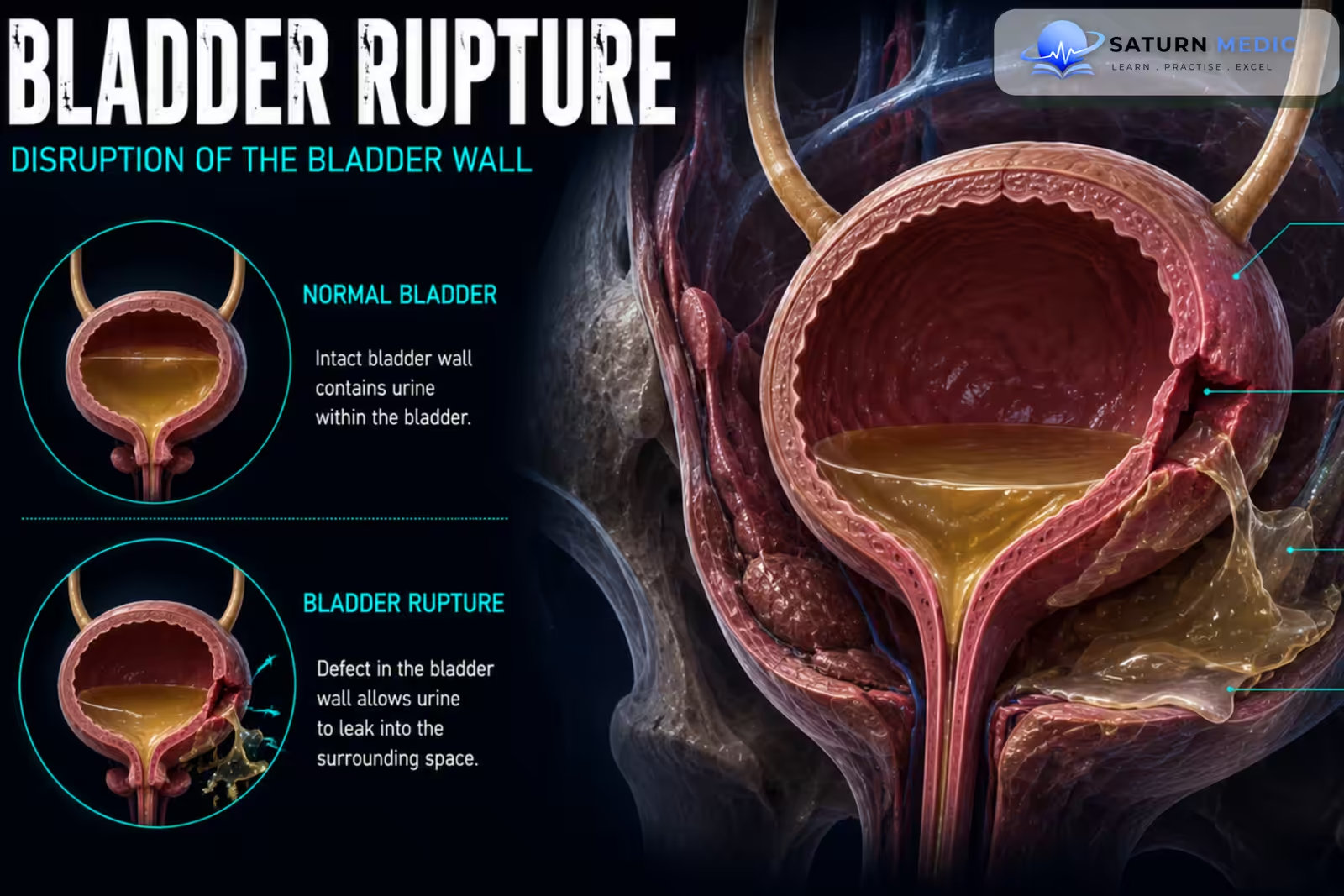
Bladder Rupture is a tear in the urinary bladder wall that allows urine, and sometimes blood, to leak into surrounding tissues or the peritoneal cavity. It usually occurs after significant trauma, particularly pelvic trauma, and requires prompt diagnosis and appropriate management.
Also called
ICD-10
Specialty
Onset
Reviewed
-
OverviewOverview
-
Etiology & Risk FactorsEtiology & Risk Factors
-
PathophysiologyPathophysiology
-
Clinical PresentationClinical Presentation
-
History TakingHistory Taking
-
Physical ExaminationPhysical Examination
-
InvestigationsInvestigations
-
DiagnosisDiagnosis
-
ManagementManagement
-
ComplicationsComplications
-
PrognosisPrognosis
-
Key Points / Clinical PearlsKey Points / Clinical Pearls
Overview
-Bladder Rupture is commonly classified into two major types:
- Extraperitoneal rupture: urine leaks into the tissues surrounding the bladder. This is the most common type and is frequently associated with pelvic fractures.
- Intraperitoneal rupture: urine leaks into the peritoneal cavity. It commonly occurs when a distended bladder ruptures at its dome and usually requires surgical repair.
-A combined injury may also occur.
Etiology & Risk Factors
-Etiology
–The main causes include:
-Traumatic Causes
- Pelvic fractures
- Road Traffic Accidents (RTA)
- Falls
- Direct lower abdominal trauma
- Penetrating pelvic injuries
-Non-Traumatic or Iatrogenic Causes
- Pelvic surgery
- Endoscopic or catheter-related injury
- Bladder overdistension
- Bladder tumor or severe inflammation in rare cases
-Risk Factors
- Pelvic fracture
- High-energy trauma
- Bladder distension at the time of injury
- Pelvic surgery
- Instrumentation of the urinary tract
Pathophysiology
-Extraperitoneal Bladder Rupture
Pelvic trauma or bone fragments → direct injury to the bladder wall → urine leaks into the extraperitoneal pelvic tissues → pelvic urinary extravasation
-Intraperitoneal Bladder Rupture
Trauma to a distended bladder → tear at the bladder dome → urine leaks into the peritoneal cavity → chemical peritonitis → peritoneal inflammation ± systemic complications
Clinical Presentation
–Symptoms
- Lower abdominal or suprapubic pain
- Difficulty passing urine
- Inability to void
- Gross hematuria
- Abdominal distension
- Pelvic pain
- Urine leakage from wounds in penetrating trauma
-Signs:
- Suprapubic tenderness
- Lower abdominal distension
- Pelvic tenderness
- Signs of associated pelvic fracture
- Reduced urine output
- Blood at the urethral meatus in associated urethral injury
History Taking
-Ask about:
- Mechanism of injury
- Pelvic or lower abdominal trauma
- Presence of a pelvic fracture
- Ability to pass urine after injury
- Gross hematuria
- Lower abdominal pain
- Time since last urination
- Previous bladder or pelvic surgery
- Recent urinary tract instrumentation
Physical Examination
-General Examination
- Airway, breathing, and circulation
- Blood pressure and heart rate
- Signs of shock
- Overall trauma assessment
-System-Specific Examination:
- Suprapubic tenderness and distension
- Pelvic tenderness or instability
- Signs of peritonitis
- External genital examination
- Check for blood at the urethral meatus
- Assess associated rectal or perineal injuries when indicated
Investigations
-Complete Blood Count
Relevant in significant trauma to assess blood loss and monitor the patient.
-Biochemistry / Specific Tests
Serum creatinine and electrolytes
Urinalysis for hematuria
Blood grouping and cross-matching in major trauma
-Imaging
CT Cystography
CT cystography is a key investigation for diagnosing Bladder Rupture in trauma patients.
It can identify:
Site of bladder rupture
Intraperitoneal or extraperitoneal urine leakage
Associated pelvic injuries
-Retrograde Cystography
A standard alternative for directly demonstrating contrast extravasation from the bladder.
-CT Abdomen and Pelvis
Useful for assessing associated abdominal and pelvic injuries.
Diagnosis
-Diagnosis of Bladder Rupture is based on:
Compatible trauma + gross hematuria or inability to void + dedicated cystography showing contrast extravasation from the bladder.
-The rupture should be classified as:
- Intraperitoneal
- Extraperitoneal
- Combined
This classification guides treatment.
Management
1. First-Line / Emergency Management
Initial management follows standard trauma principles:
Stabilize Airway, Breathing, and Circulation
Treat associated life-threatening injuries
Provide analgesia
Establish urinary drainage when safe and appropriate
Urgent trauma and urological assessment
If urethral injury is suspected, evaluate the urethra before attempting urethral catheterization.
2. Definitive Treatment
Treatment depends mainly on the type and complexity of rupture.
Uncomplicated extraperitoneal rupture: usually managed with continuous bladder drainage.
Intraperitoneal rupture: usually requires surgical repair.
Complicated extraperitoneal rupture: may require surgical repair.
3. Medical Treatment
Analgesia
Fluid resuscitation when required
Antibiotics when clinically indicated
Correction of electrolyte abnormalities
Treatment of associated infection or sepsis
4. Surgical / Procedural Treatment
Extraperitoneal Bladder Rupture
Usually managed with:
Urethral catheter drainage for an appropriate healing period
Follow-up cystography before catheter removal when indicated
–Surgical repair may be required if there is:
Bladder neck involvement
Associated rectal or vaginal injury
Bone fragments within the bladder
Persistent urine leakage
Injury requiring open pelvic surgery
-Intraperitoneal Bladder Rupture
Usually requires:
Surgical exploration
Primary repair of the bladder
Postoperative bladder drainage
5. Supportive Management
Monitor urine output
Monitor renal function and electrolytes
Treat associated pelvic injuries
Complications
- Urinary leakage
- Urinoma
- Urinary ascites
- Peritonitis
- Sepsis
- Pelvic abscess
- Acute Kidney Injury (AKI)
- Electrolyte abnormalities
- Persistent fistula
- Bladder dysfunction
- Urinary incontinence
- Urethral stricture
Prognosis
-The prognosis is generally good when Bladder Rupture is diagnosed promptly and managed appropriately.
-Uncomplicated extraperitoneal injuries usually heal with bladder drainage.
-Intraperitoneal injuries have a higher risk of peritonitis and systemic complications if treatment is delayed but generally have good outcomes after timely surgical repair.
-Prognosis is also influenced by the severity of associated pelvic and abdominal injuries
Key Points / Clinical Pearls
- Bladder Rupture is usually caused by significant pelvic or lower abdominal trauma.
- Pelvic fractures are strongly associated with extraperitoneal bladder injury.
- Gross hematuria is an important warning sign.
- Difficulty voiding and suprapubic pain may occur.
- Extraperitoneal rupture is the most common type.
- Intraperitoneal rupture usually occurs at the bladder dome.
- A distended bladder is more vulnerable to intraperitoneal rupture.
- Dedicated CT cystography or retrograde cystography confirms the diagnosis.
- Routine CT without proper bladder filling may miss the injury.
- Associated urethral injury should be considered before urethral catheterization.
- Uncomplicated extraperitoneal rupture is usually treated with catheter drainage.
- Intraperitoneal rupture usually requires surgical repair.
- European Association of Urology (EAU). EAU Guidelines on Urological Trauma .
- Morey AF, Brandes S, Dugi DD III, et al. Urotrauma: AUA Guideline. J Urol. 2014;192(2):327-335. Journal of Urology .
- American Urological Association (AUA). Urotrauma Guideline .
- Gomez RG, Ceballos L, Coburn M, et al. Consensus Statement on Bladder Injuries. BJU Int. 2004;94(1):27-32. PubMed .
- Phillips B, Holzmer S, Turco L, et al. Trauma to the Bladder and Urethra: A Review of Diagnosis and Management. J Trauma Acute Care Surg. 2017;83(5):S144-S149.
- National Library of Medicine (NIH). Bladder Trauma . StatPearls.