

Clinical Subject Page
Hypersensitivity pneumonitis (HP)
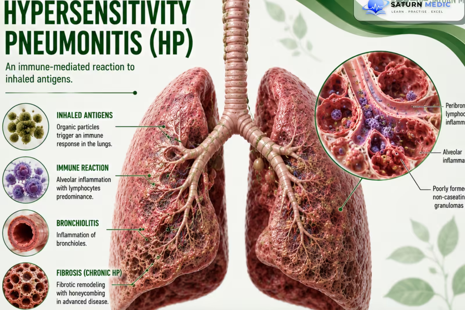
Hypersensitivity pneumonitis (HP) is an immune-mediated inflammatory lung disease caused by repeated inhalation of an environmental antigen in a susceptible person.
Also called
ICD-10
Specialty
Onset
Reviewed
-
OverviewOverview
-
Etiology & Risk FactorsEtiology & Risk Factors
-
PathophysiologyPathophysiology
-
Clinical PresentationClinical Presentation
-
History TakingHistory Taking
-
Physical ExaminationPhysical Examination
-
InvestigationsInvestigations
-
DiagnosisDiagnosis
-
ManagementManagement
-
ComplicationsComplications
-
PrognosisPrognosis
-
Key Points / Clinical PearlsKey Points / Clinical Pearls
Overview
Hypersensitivity pneumonitis (HP) occurs when inhaled antigens trigger an abnormal immune response in the:
- Alveoli
- Small airways
- Lung interstitium
Common sources include:
- Birds
- Mold
- Fungi
- Agricultural dusts
- Contaminated water or humidifiers
Repeated exposure may cause:
Inflammation → Lung injury → Fibrosis
Etiology & Risk Factors
Common Causes of Hypersensitivity pneumonitis (HP)
-Bird Exposure
Bird feathers
Bird droppings
Bird proteins
-Often called bird fancier’s lung.
Moldy hay
Damp buildings
Compost
Contaminated ventilation systems
-Agricultural Exposure
Hay
Grain
Farming dusts
Often associated with farmer’s lung.
-Other Exposures
Humidifiers
Hot tubs
Contaminated water systems
Certain occupational chemicals
-Risk Factors
Repeated antigen exposure
High-intensity exposure
Farming
Bird keeping
Working with mold or organic dust
Certain occupational environments
Genetic susceptibility
Pathophysiology
Inhalation of environmental antigen
⬇
Antigen reaches the small airways and alveoli
⬇
Abnormal immune response
⬇
Lymphocytic inflammation
⬇
Small airway + Interstitial lung injury
⬇
Granuloma formation in some patients
⬇
Repeated or persistent exposure
⬇
Chronic inflammation
⬇
Pulmonary fibrosis
-Key Concept
Early inflammatory disease may improve after antigen avoidance, while established fibrosis may be irreversible
Clinical Presentation
Acute Features of Hypersensitivity pneumonitis (HP)
Symptoms may develop within hours after heavy exposure:
Fever
Chills
Dry cough
Dyspnea
Fatigue
Malaise
Symptoms may improve after leaving the exposure.
-Chronic Features of Hypersensitivity pneumonitis (HP)
Progressive exertional dyspnea
Persistent cough
Fatigue
Reduced exercise tolerance
Weight loss
Signs
Tachypnea
Fine inspiratory crackles
Reduced oxygen saturation
-Advanced Disease
Digital clubbing
Cyanosis
Signs of pulmonary hypertension
Signs of respiratory failure
History Taking
- Shortness of breath?
- Dry cough?
- Fever or chills after exposure?
- When did symptoms start?
- Do symptoms improve away from home or work?
- Bird exposure?
- Feather pillows or bedding?
- Mold or dampness at home?
- Farming or hay exposure?
- Hot tub use?
- Humidifier use?
- Occupational exposure?
Physical Examination
-General Examination
Look for:
- Tachypnea
- Respiratory distress
- Reduced oxygen saturation
- Cyanosis
-Chest Examination
May show:
- Fine inspiratory crackles
- Inspiratory squeaks
-Chronic or Advanced Disease
Look for:
-
- Digital clubbing
- Signs of pulmonary hypertension
- Signs of cor pulmonale
Investigations of Hypersensitivity pneumonitis (HP)
-High-Resolution CT (HRCT) Chest — Key Imaging Test
May show:
-
Ground-glass opacities
-
Centrilobular nodules
-
Mosaic attenuation
-
Air trapping
-
Fibrosis in chronic disease
-Important Imaging Feature
The combination of:
-
Ground-glass changes
-
Mosaic attenuation
-
Air trapping
supports the diagnosis of HP.
-Pulmonary Function Tests
May show:
-
Restrictive pattern
-
Obstructive pattern
-
Mixed pattern
-
Reduced DLCO
-Blood Tests
May include:
-
Serum antigen-specific IgG antibodies
-
Inflammatory markers
-Important Note
Positive antigen-specific IgG indicates exposure but does not alone confirm the diagnosis.
-Bronchoalveolar Lavage (BAL)
May show:
-
Increased lymphocytes
-Lung Biopsy
Consider when:
-
-
Diagnosis remains uncertain
-
Clinical and imaging findings are inconclusive
-
Diagnosis of Hypersensitivity pneumonitis (HP)
Hypersensitivity Pneumonitis · Diagnostic Confidence Levels & Stepwise Workup — ATS 2020
CD4:CD8 ratio <1 (inverted — opposite to sarcoidosis); BAL mast cells may be elevated
Related Topics
Management Hypersensitivity pneumonitis (HP)
-Main Goals of Management of Hypersensitivity pneumonitis (HP)
-
Identify the responsible antigen
-
Stop further exposure
-
Reduce lung inflammation
-
Prevent pulmonary fibrosis
-Antigen Avoidance — Most Important Treatment
-
Remove the responsible exposure
-
Improve ventilation
-
Remove mold contamination
-
Avoid responsible birds or animals
-
Use appropriate workplace protection
Important Note
Complete antigen avoidance is the cornerstone of treatment.
-Corticosteroids
May be used in patients with:
-
Significant symptoms
-
Severe inflammatory disease
-
Progressive disease
They may improve symptoms and speed recovery but do not replace antigen avoidance.
-Fibrotic Disease
Management may include:
-
-
Treatment of progressive pulmonary fibrosis
-
Antifibrotic therapy in selected patients
-
Complications
- Chronic pulmonary fibrosis
- Progressive loss of lung function
- Chronic hypoxemia
- Respiratory failure
- Pulmonary hypertension
- Cor pulmonale
- Acute exacerbation
- Death
Prognosis of Hypersensitivity pneumonitis (HP)
- Prognosis depends mainly on:
- Early identification of the antigen
- Successful antigen avoidance
- Presence or absence of fibrosis
- Severity of lung impairment
- Nonfibrotic HP may improve significantly after exposure stops.
- Fibrotic HP may continue to progress despite antigen avoidance.
- Established pulmonary fibrosis is associated with a worse prognosis.
- Early diagnosis and complete exposure avoidance improve outcomes.
Key Points / Clinical Pearls of Hypersensitivity pneumonitis (HP)
- Hypersensitivity pneumonitis (HP) is an immune-mediated lung disease caused by inhaled environmental antigens.
- Birds, mold, and farming exposures are common causes.
- A detailed exposure history is essential.
- Symptoms may worsen after exposure and improve when away from it.
- HRCT is the key imaging test.
- BAL often shows lymphocytosis.
- Positive antigen-specific IgG indicates exposure, not necessarily disease.
- Sirajuddin A, Kanne JP. National Center for Biotechnology Information (NIH). Hypersensitivity Pneumonitis, StatPearls.
- MedlinePlus, National Library of Medicine (NIH). Hypersensitivity Pneumonitis: Medical Encyclopedia.
- Raghu G, Remy-Jardin M, Ryerson CJ, et al. Diagnosis of Hypersensitivity Pneumonitis in Adults: An Official ATS/JRS/ALAT Clinical Practice Guideline. Am J Respir Crit Care Med. 2020. PMID: 32706311.